Vertucci configurations, canal number and shape by tooth group, clinically significant variations
ICD-10: —
Root Canal System: Why "System" and Not "Canal"
A 34-year-old patient returns eight months after endodontic treatment of tooth 16. The follow-up radiograph shows two canals filled to the apex and a third filled almost to the apex — the picture looks acceptable. Yet the tooth is tender to biting and a radiolucency has appeared at the apex of the mesial root. On retreatment under magnification a fourth, untreated canal is found — the second mesiobuccal canal (MB2). The error was not made during obturation; it was made at the level of anatomical expectation. The clinician treated three canals because three were expected.
This is the typical failure scenario. Instruments, irrigants and materials only act within the space the clinician has located. Everything not located remains a reservoir of infection. Endodontics therefore begins with anatomy, not with a file.
General tooth structure — enamel, dentin, pulp, cementum — is already familiar (Propaedeutics course, module p1-m4), and the three-dimensional topography of the roots was explored on the model in module e1-m1. This article is the text base for test e1-m2: it systematises the number, shape and configuration of canals for every tooth group and provides the vocabulary used to describe that anatomy.
MB2 in maxillary first molars: 55–95% — across studies; 60–70% most often in clinical practice
Maxillary first molar: 3 roots / 4 canals — the typical norm, not a variation
Apical constriction: 0.5–1.0 mm — short of the radiographic apex; diameter 0.2–0.3 mm
Isthmus, mesial root of 36 / 46: over half of roots — 3–5 mm from the apex (von Arx, 2005)
Incisors and canines: 1 root / 1 canal — second canal in mandibular incisors ≈ 10–41%
Maxillary first premolar: 2 canals ≈ 85–95% — yet two roots in only 55–60%
The root canal system is the entire internal space of the root lined by pulp or its remnants: the main canals, lateral and accessory canals, apical ramifications (apical delta), isthmuses and transverse anastomoses between canals.
A useful analogy is not a water pipe but a river system: one main channel, side branches, connections between them and a fan of distributaries at the mouth. A pipe can be mechanically cleaned along its whole length; a river network cannot. This is precisely why canal preparation is called chemo-mechanical — the instrument travels along the main channel while the irrigant must reach everything else.
Two practical consequences follow from the definition: the number of canals does not equal the number of roots, and canal configuration is described not as present/absent but as a branching type. The following sections develop both statements.
Landmarks: From the Orifice to the Apical Foramen
Anatomy can only be described through fixed reference points. Endodontics uses two groups of them: coronal — the canal orifice and the pulp chamber floor, where the search begins; and apical — the constriction and the foramina at which the canal ends. Everything else lies in between: the apical delta, lateral canals, isthmuses. These words are used as working terms throughout the article, so they are defined here, before the tooth groups.
The orifice is the entrance to a canal on the pulp chamber floor. Its position is not random: Krasner and Rankow (J Endod, 2004) described the architecture of the chamber floor and reduced it to nine regularities known as the laws of the pulp chamber floor. Five of them carry most of the clinical weight. The law of centrality: the floor of the pulp chamber is located at the center of the tooth at the level of the cementoenamel junction. The law of concentricity: at that level the external outline of the crown reproduces the outline of the chamber — crown shape predicts floor shape. The law of the CEJ: the cementoenamel junction is the most consistent landmark for locating the pulp chamber. The laws of symmetry: the orifices are equidistant from a line drawn mesiodistally through the center of the floor and lie on a perpendicular to that line — both laws fail for maxillary molars, which is exactly why MB2 has to be sought separately. The law of color change and the laws of orifice location: the chamber floor is always darker than the walls, and an orifice always lies at the junction of wall and floor, at the angles of that junction and at the terminus of a root fusion (developmental) line. The practical point is that an orifice is not hunted at random but expected in a predictable place.
The apical landmarks are more difficult, because some of them are not visible on a radiograph at all and require magnification or CBCT (cone-beam computed tomography). The key one is the apical constriction: the narrowest point of the canal, the level to which preparation and obturation are carried. The apical constriction lies 0.5–1.0 mm short of the radiographic apex, and it is from this rule that working length is derived in module e2-m1 of the course "Instrumental Canal Processing". The diameter of the constriction is of the order of 0.2–0.3 mm; with age it shifts and loses definition because of cementum apposition (Kuttler, 1955).
Orifice Canal orifice — The funnel-shaped entrance to a canal on the pulp chamber floor. The only landmark of the canal system accessible to direct inspection, and the point at which mechanical preparation begins. The task of the access stage is to find EVERY orifice: a missed orifice means a missed canal, however good the subsequent work.
Constriction Apical constriction (minor foramen) — The narrowest point of the canal, approximately coinciding with the cementodentinal junction. It lies 0.5–1.0 mm short of the radiographic apex and measures of the order of 0.2–0.3 mm in diameter. This is the physiologic limit of preparation and obturation; with age it migrates apically and becomes less distinct (Kuttler, 1955).
Foramina Physiologic and anatomic apical foramen — The physiologic foramen is the canal exit at the level of the apical constriction — the target the clinician works to. The anatomic foramen (major foramen) is the actual opening on the root surface; it lies further apically and frequently on the side of the apex rather than at its tip. Working "to the anatomic foramen" means working beyond the canal, into the periodontium.
Apex Radiographic apex — The most apical point of the root shadow on a radiograph. It is a measuring reference, not an anatomic boundary: the anatomic foramen usually does not coincide with it, and the position of the apex itself depends on projection. Hence the 0.5–1.0 mm rule rather than working "to the apex".
Delta / lateral Apical delta, lateral and accessory canals — The apical delta is a fan of small ramifications of the main canal in the apical third instead of a single exit. Lateral canals branch off the main canal at an angle in the middle third of the root, accessory canals in the apical third; both connect the canal with the periodontium. Neither can be negotiated with an instrument, so both belong to the irrigant; on a periapical radiograph they are mostly visible only once filled with sealer or gutta-percha.
Isthmus Isthmus — A narrow slit-like connection between two canals of the same root, containing pulp or its remnants. In the mesial root of mandibular molars an isthmus is found 3–5 mm from the apex in more than half of roots (von Arx, Int Endod J, 2005); isthmus types I–V — from a simple connecting strand to a complete corridor across the whole section — come from the Hsu and Kim classification (1997). It cannot be negotiated with a round instrument.
Canal Configurations: Vertucci and Weine
Canal number is only half the description. One and the same root with two canals may exit through a single foramen or through two, and the two canals may merge in mid-root and separate again; clinically these are different tasks. Root anatomy is therefore described not by a count but by a branching type. Vertucci (1984; reviewed in 2005) examined cleared extracted teeth and defined eight configuration types, each conveniently written as a formula of "number of orifices — number of foramina": 1-1, 2-1, 1-2, 2-2 and so on. The essential caveat, and the commonest source of confusion: a type describes ONE root, not the whole tooth.
A shorter classification by Weine is used alongside Vertucci. Its first version (1969) described three configurations of the mesiobuccal root of tooth 16; a fourth type was added in later editions of the textbook "Endodontic Therapy". The four Weine types are convenient for quickly describing a root with two canals, but they do not cover the intermediate Vertucci patterns — hence the "no equivalent" entries in the correspondence table below.
The practical significance of the type lies not at the orifice-location stage but in the apical third: it determines how many foramina must be sealed and whether the canals have to be prepared independently of each other. The type cannot be established from a single periapical radiograph — it emerges during canal negotiation or from CBCT.
I (1-1) Type I — a single canal throughout — One canal runs from the orifice to a single apical foramen. The simplest and most predictable configuration. Typical examples — maxillary incisors and canines, the palatal root of the maxillary first molar.
II (2-1) Type II — two canals merging into one — Two separate canals leave the pulp chamber, merge within the root and exit through a single foramen. Coronally it looks exactly like type IV — two orifices — so the second orifice is just as easy to miss. The classic example is the mesiobuccal root of the maxillary first molar, in which MB2 merges with MB1.
III (1-1-1) Type III — one canal divides and rejoins — One canal leaves the chamber, divides into two within the root and rejoins into one before the foramen. One orifice, one foramen, and two lumina in the middle third — a pattern a radiograph cannot show. Described in mandibular incisors and premolars.
IV (2-2) Type IV — two separate canals — Two canals run separately from their orifices to two distinct foramina. Each requires independent preparation and obturation. Examples — the mesial root of the mandibular first molar, and a single-rooted maxillary first premolar with two canals.
V (1-2) Type V — the canal divides in the apical third — One canal leaves the chamber and divides in the apical third into two, each with its own foramen. The most treacherous configuration to prepare: there is only one orifice, so the second canal is easily overlooked, and it appears exactly where visibility is worst. Most characteristic of mandibular premolars.
VI (2-1-2) Type VI — two canals merge and separate again — Two canals leave the chamber, join in the middle third of the root and separate again towards two foramina. Rare; clinically it shows itself when an instrument entered through one orifice emerges from the "wrong" foramen.
VII (1-2-1-2) Type VII — double division — One canal divides, rejoins, and in the apical third divides finally into two foramina. The most demanding of the rare configurations to prepare mechanically. Described mainly in mandibular incisors and premolars.
VIII (3-3) Type VIII — three separate canals — Three separate canals run within one root from their orifices to three foramina. The principal example is the mesial root of mandibular molars (MB, middle mesial and ML); the second most frequent is the mesiobuccal root of maxillary molars.
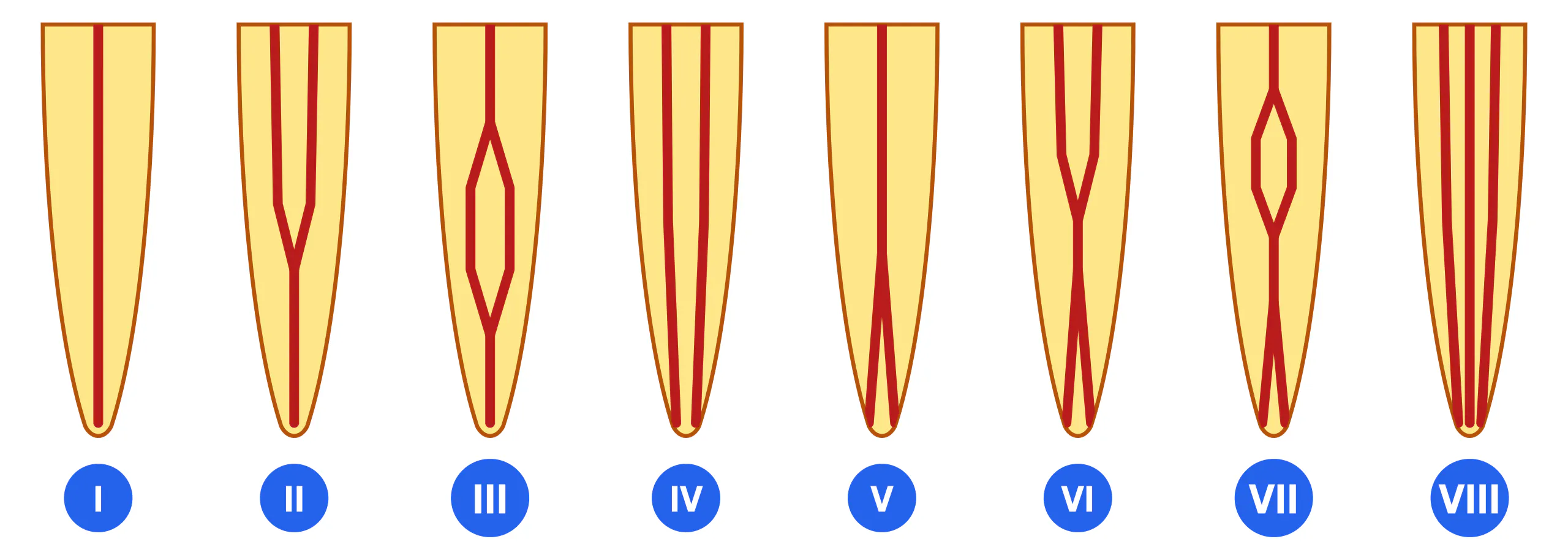
| Vertucci type (1984) | Structure | Weine (1969, extended in later editions) |
|---|---|---|
| I (1-1) | One canal from orifice to a single foramen | Type I |
| II (2-1) | Two canals merge before the apex, one foramen | Type II |
| III (1-1-1) | The canal divides within the root and rejoins into one | No equivalent |
| IV (2-2) | Two separate canals, two foramina | Type III (2-2) |
| V (1-2) | One canal divides in the apical third, two foramina | Type IV (1-2) |
| VI (2-1-2) | Two canals merge in the middle third and separate again | No equivalent |
| VII (1-2-1-2) | The canal divides, rejoins and divides again | No equivalent |
| VIII (3-3) | Three separate canals, three foramina | No equivalent |
Incisors and Canines: Number, Shape, Configuration
The anterior group is the simplest in terms of root number (always one root) and at the same time a source of avoidable failures because of canal cross-sectional shape.
Maxillary incisors and canines: the canal is wide, round or oval in cross-section, and the configuration is almost always type I. The maxillary canine is the longest tooth in the arch (average of the order of 26–27 mm — mean lengths after Wheeler's Dental Anatomy, Physiology and Occlusion / Nelson), which matters when selecting instrument length.
Mandibular incisors: the root is compressed mesiodistally and the canal is ribbon-shaped in cross-section. Such a slit readily separates into buccal and lingual portions, hence the high proportion of second canals. A second canal in mandibular incisors is reported in approximately 10–41% of cases across samples (Vertucci, 1984 — about 30%, as types II, III and V; the upper bound comes from Benjamin and Dowson, 1974), whereas two separate apical foramina occur in fewer than 5%. In other words, a second canal is usually present but exits together with the first.
| Tooth (FDI) | Roots | Canals — typical | Cross-section | Variations and frequency |
|---|---|---|---|---|
| 11 / 21 — central incisor | 1 | 1 | Round, wide | Type I in virtually 100%; two canals are a curiosity |
| 12 / 22 — lateral incisor | 1 | 1 | Oval | Type I in virtually 100%; the tooth most often affected by dens invaginatus (a developmental anomaly: invagination of enamel and dentin into the tooth; detailed in the "Variations" section) |
| 13 / 23 — canine | 1 | 1 | Oval, elongated buccolingually | Type I in virtually 100%; two canals are isolated reports |
| 31 / 41 — central incisor | 1 | 1 (less often 2) | Ribbon-shaped | Second canal ≈ 10–41% across samples; two separate foramina < 5% |
| 32 / 42 — lateral incisor | 1 | 1 (less often 2) | Ribbon-shaped | Frequencies close to the central incisor; the additional canal is lingual |
| 33 / 43 — canine | 1 | 1 | Oval, often flattened | Two canals in roughly 10–20%; two roots are rare |
Premolars: The Group with the Widest Anatomical Range
Premolars show the widest anatomical variation relative to their modest size. The maxillary first premolar is the only tooth outside the molar region for which two roots are the norm rather than a variation; three roots occur occasionally. Mandibular premolars are nominally single-rooted, yet they divide the canal in the apical third (type V) more often than any other tooth, and they are a frequent source of missed canals and perforations among inexperienced clinicians.
It is on the maxillary first premolar that the difference between root number and canal number is clearest. Two roots — buccal and palatal — are present in approximately 55–60% of maxillary first premolars; the remainder have one root, occasionally three. Two canals, however, are present in a substantially larger share of teeth, of the order of 85–95%: a single-rooted maxillary first premolar very often carries two canals within that single root (Vertucci types II, IV or V). "One root" on a radiograph therefore does not mean "one canal" (Vertucci, 1984; Vertucci, 2005; Cohen's Pathways of the Pulp, 12th ed.). Three-rooted variants are a small but clinically important minority, since the third (palatal) root is easily missed if the radiograph is not assessed in two projections.
A note on the figures encountered in the literature and in teaching materials. A value of about 60–70% for the maxillary first premolar describes the proportion of TWO-ROOTED teeth (reported closer to 55–60%), not of two-canalled ones. This is the tooth on which root number and canal number are confused most often, so whenever a premolar figure is quoted, establish first whether it refers to roots or to canals.
A distinctive feature of the mandibular first premolar is the radicular groove on the lingual or mesial surface of the root. It corresponds to internal division of the canal and serves as a landmark: if the groove is present, expect a second canal.
| Tooth (FDI) | Roots | Canals — typical | Variations and frequency |
|---|---|---|---|
| 14 / 24 — first premolar | 2 in ≈ 55–60%, sometimes 1, rarely 3 | 2 | Two canals ≈ 85–95% — frequently within a single root as well; one canal ≈ 5–10%; three roots with three canals ≈ 1–6%. The 55–60% figure is the proportion of two-rooted, not two-canalled, teeth |
| 15 / 25 — second premolar | 1 in most cases | 1–2 | One canal in roughly half of teeth, two in the remainder; reported figures vary widely (Vertucci, 1984: type I in about half of specimens). Three canals are rare |
| 34 / 44 — first premolar | 1 | 1 (less often 2 or more) | One canal ≈ 70–75%; apical-third division (type V) and other patterns ≈ 25–30%. Landmark: the radicular groove |
| 35 / 45 — second premolar | 1 | 1 | One canal in the great majority of cases (of the order of 85–97% across samples); two canals substantially less often than in the first premolar |
Molars: The Highest Canal Count and the Highest Risk of a Missed Canal
Molars carry the highest number of canals and account for the highest number of missed ones. The vocabulary here is dense, so it is fixed at the outset: canals are named after their root and side — mesiobuccal (MB), distobuccal (DB) and palatal (P) in maxillary molars; mesiobuccal (MB), mesiolingual (ML) and distal (D) in mandibular molars. The first and second canals of the mesiobuccal root of a maxillary molar are designated MB1 and MB2.
The maxillary first molar is the reference example of the mismatch between root number and canal number: three roots and, as a rule, four canals. The second mesiobuccal canal (MB2) is found in 55–95% of cases across studies, and 60–70% most often in clinical practice; it leaves MB1 mesiopalatally and is frequently covered by overhanging dentin. The most curved and least accessible root is the mesiobuccal, and within it MB2 is more curved than MB1. The palatal root of 16 / 26, by contrast, is longer than the buccal roots, usually straight and wide, and therefore traceable on a periapical radiograph better than the others; it almost always contains a single canal. In the maxillary second molar the roots are more often approximated or fused, and MB2 is found less often — of the order of 20–40% of cases.
The mandibular first molar: two roots and most often three canals — two in the mesial root (MB and ML) and one in the distal. The distal root of 36 / 46 contains two canals in roughly one third of teeth, so four canals in this tooth are not unusual. There is almost always an isthmus between the mesial canals, and a middle mesial canal may open within its groove; no single prevalence figure exists in the literature: clinical studies using an operating microscope report figures in the region of 20% (Karapinar-Kazandag, Basrani, Friedman, J Endod, 2010), micro-CT data and young samples report considerably more, and older radiographic work reported only a few per cent. A separate variation is radix entomolaris, an additional distolingual root: 3–5% in European samples and up to 30–40% in East Asian ones; the clinical approach to it was systematised by Calberson, De Moor and Deroose (2007), while an earlier clinically oriented description was published by De Moor, Deroose and Calberson (Int Endod J, 2004). The mandibular second molar more often has approximated or fused roots, the extreme expression of which is the C-shaped configuration: 3–8% in European samples, 30–45% in East Asian populations, while pooled data without separation by population give 8–44% (Fan et al., 2004; categories C1–C5). The letter C in the name refers to the shape of the canal in cross-section, not to an alphabetical label. Third molars stand apart: root number, degree of fusion, canal number and canal course are all unpredictable, no typical norm exists for them, and unpredictable anatomy is a typical justified indication for CBCT.
| Tooth (FDI) | Roots | Canals — typical | Variations and frequency |
|---|---|---|---|
| 16 / 26 — first molar | 3: MB, DB, palatal | 4 (less often 3) | MB2 — 55–95% across studies, 60–70% most often in clinical practice; the palatal root almost always has one canal; two canals in DB and root fusion are rare |
| 17 / 27 — second molar | 3, not infrequently approximated or fused | 3 (less often 4) | MB2 of the order of 20–40%; root fusion and single-canal fused variants are more frequent than in 16 / 26 |
| 18 / 28 — third molar | Inconsistent: 1–3, often fused | Inconsistent: 1–4 | No typical norm exists, canal number and course are unpredictable; unpredictable anatomy is a typical justified indication for CBCT |
| 36 / 46 — first molar | 2: mesial, distal; occasionally 3 — radix entomolaris | 3 (less often 4) | Two canals in the mesial root (MB, ML) almost always, with an isthmus between them in more than half of roots; the distal root has two canals in roughly one third of teeth; middle mesial canal in the region of 20% when working with a microscope; radix entomolaris 3–5% (Europe) to 30–40% (East Asia) |
| 37 / 47 — second molar | 2, often fused | 3 (less often 2) | C-shaped configuration: 3–8% in European samples, 30–45% in East Asian populations; pooled data without separation by population give 8–44% (Fan et al., 2004; categories C1–C5) |
| 38 / 48 — third molar | Inconsistent: 1–3, often fused into a conical root | Inconsistent: 1–4 | No typical norm exists; root fusion and C-shaped configurations are frequent; the justification for CBCT is the same as for 18 / 28 |
The Variations Most Often Missed
MB2 Second mesiobuccal canal of maxillary molars — The second canal of the mesiobuccal root. It often leaves MB1 at an acute angle and merges with it near the apex (a type II configuration), so its absence on a radiograph proves nothing. One of the leading causes of failure in maxillary molar endodontics.
MMC Middle mesial canal of mandibular molars — An additional canal in the groove between the MB and ML orifices of the mesial root, frequently an opened isthmus. No single prevalence figure exists: clinical studies using the operating microscope report figures in the region of 20% (Karapinar-Kazandag, Basrani and Friedman, J Endod, 2010), studies on young patients and micro-CT series report considerably more, and older radiographic work reported only a few per cent. The probability of finding it is higher in young patients.
RE / RP Radix entomolaris and radix paramolaris — An additional root of a mandibular molar: entomolaris is distolingual, paramolaris is mesiobuccal. The root is usually slender and curved lingually. The clinical approach to such a root was systematised by Calberson, De Moor and Deroose (J Endod, 2007); an earlier clinically oriented description was published by De Moor, Deroose and Calberson (Int Endod J, 2004). Frequency: 3–5% in European samples, up to 30–40% in East Asian populations.
C-shaped C-shaped configuration — A result of root fusion (most often mesial and distal roots of the mandibular second molar): instead of separate orifices there is a continuous or interrupted C-shaped arc. Classifications: Melton et al. (J Endod, 1991) — types I–III by sectional shape; Fan et al. (J Endod, 2004) — refined categories C1–C5. The problem with this anatomy is not locating it but the impossibility of mechanically preparing the whole arc with a round instrument.
Isthmus Isthmus — A narrow slit-like connection between two canals of the same root, containing pulp or its remnants. In the mesial root of the mandibular first molar an isthmus is found 3–5 mm from the apex in more than half of roots (von Arx, Int Endod J, 2005); the isthmus types themselves come from the Hsu and Kim classification (1997). It cannot be negotiated with an instrument and is cleaned only by irrigation and its activation.
Dens invaginatus Dens invaginatus — A developmental anomaly: invagination of enamel and dentin into the tooth before eruption, creating an additional cavity communicating with the oral environment. Most characteristic of the maxillary lateral incisor. The canal system in such a tooth differs fundamentally from the typical pattern — one of the most self-evident justified indications for CBCT at the planning stage.
| Variation | Where to look | Confirmed by | Reported frequency |
|---|---|---|---|
| MB2 (16 / 26) | Mesiopalatal to MB1, on the MB1–palatal orifice line | Magnification, probing beneath overhanging dentin, CBCT | 55–95% depending on method; 60–70% in clinical studies |
| Middle mesial canal (36 / 46) | The isthmus groove between the MB and ML orifices | Operating microscope, CBCT, micro-CT (laboratory) | Data diverge: from a few per cent to ≈ 20% and above; more often in young patients |
| Radix entomolaris | Additional distolingual root; orifice distolingual | Angled radiograph, CBCT | 3–5% (European samples), up to 30–40% (East Asia) |
| C-shaped configuration (37 / 47) | Chamber floor: a continuous arc instead of separate orifices | CBCT, inspection of the floor under magnification | 3–8% (European samples), 30–45% (East Asia); pooled 8–44% |
| Mesial root isthmus | Between MB and ML, 3–5 mm from the apex | Magnification; micro-CT; at surgery — the resected root face | Over half of roots in the apical third (von Arx, 2005; isthmus types — Hsu, Kim, 1997) |
| Second canal of a mandibular incisor | Lingual to the main canal, within the ribbon-shaped cross-section | Angled radiograph, CBCT, magnification | ≈ 10–41% across samples; two foramina < 5% |
Canal Curvature and Age-Related Changes
Straight canals barely exist. Curvature in the mesiodistal plane is visible on a radiograph; curvature in the buccolingual plane is not visible at all and is therefore systematically underestimated.
The standard descriptor is the Schneider angle (Schneider, 1971): a line is drawn along the coronal part of the canal, a second line is drawn from the point where the canal begins to deviate to the apical foramen, and the angle between them expresses the degree of curvature. The method is convenient but says nothing about how abruptly the turn occurs, so it is complemented by the radius of curvature (Pruett et al., 1997): for the same angle a small radius means a sharp bend over a short distance — and a greater load on the instrument. A note on the grades listed below: category boundaries are only approximate in the literature, which is why the intervals 5–10° and 20–25° are not named — they fall on the border between categories, where no hard threshold exists.
Practical guide by group: curvature is most pronounced in the mesiobuccal root of the maxillary first molar and in the mesial canals of mandibular molars; the palatal root of a maxillary molar is by contrast usually straight and wide; the distal root of a mandibular molar is moderately curved. Curvature cannot be fully described from a single radiograph — two projections or CBCT are required.
≤ 5° Straight canal — Schneider angle not exceeding 5°. The most technically predictable anatomy. Typical example — the palatal root of the maxillary first molar.
10–20° Moderate curvature — Angle in the range of about 10–20°. Must be accounted for when selecting instruments but remains manageable. Example — the distal root of the mandibular first molar.
≥ 25° Severe curvature — Angle of 25° and above. The highest risk of shaping errors. Examples — the mesiobuccal root of the maxillary first molar, the mesial canals of mandibular molars, radix entomolaris.
S S-shaped (bayonet) canal — Double curvature: a bend in one direction in the middle portion and in the opposite direction apically. A single Schneider angle does not describe it; two measurements are needed. Most often described in premolars and in the mesiobuccal root of maxillary molars.
The canal system is not static — it narrows throughout life. Odontoblasts continuously deposit secondary dentin, so the pulp chamber shrinks, the orifices shift and become less conspicuous, and the canals narrow, coronally first. Caries, attrition and trauma induce local tertiary (reparative) dentin, and after trauma calcific metamorphosis may occur — rapid obliteration of the lumen over a considerable length.
Pulp stones (denticles) are discrete calcifications within the chamber; the reported detection rate depends heavily on method — radiographic studies find them in a proportion of patients (published figures differ severalfold), whereas histological examination detects them far more often. What matters clinically is not the count but the consequence: a pulp stone over an orifice can occlude the canal entrance completely.
At the apex cementum apposition continues, displacing the apical foramen further from the anatomic apex and making the apical constriction less distinct (described by Kuttler as early as 1955). Consequence: in an elderly patient the landmarks of the apical third are less reliable than in a young one, for an identical radiographic appearance.
How Canal Anatomy Is Assessed Before and During Treatment
All of the variations above are useless as knowledge unless the clinician knows which method reveals each of them. Below is what every method contributes specifically to assessment of canal system anatomy. Techniques of performance belong to other modules: working length determination is covered in module e2-m1 of the course "Instrumental Canal Processing", and radiographic interpretation of pathology in the radiology course.
| Method | Finding | Note |
|---|---|---|
| Periapical radiograph (single projection) | Root number, approximate length, mesiodistal curvature, signs of obliteration | Canals lying one behind the other (buccal behind lingual) superimpose and appear as one — the method systematically underestimates canal number |
| Angled radiograph, 20–30° tube shift | Separation of superimposed canals, the second incisor canal, an additional mandibular molar root | SLOB rule (Same Lingual, Opposite Buccal): when the source is shifted, the image of a lingual object moves in the same direction and a buccal object in the opposite direction |
| CBCT (cone-beam computed tomography) | Three-dimensional assessment of root and canal number, configuration type, curvature in both planes, C-shaped anatomy, additional roots | Prescribed on justified indications when complex anatomy is suspected, not routinely (AAE and AAOMR joint position statement on CBCT in endodontics) |
| Magnification: loupes ×2.5–×6, operating microscope up to ×25 | Orifices hidden by overhanging dentin, isthmuses, colour change of the chamber floor, cracks | The difference in MB2 detection rates between studies with and without magnification is a direct consequence of this method |
| Electronic apex locator | Confirmation that a located canal is patent and identification of the apical constriction level | For anatomy it serves as a confirmatory tool: the device response distinguishes a true canal from a depression in dentin |
| Micro-CT (micro-focus computed tomography) | Reference-standard visualisation of the canal system including isthmuses and the smallest ramifications | A laboratory method on extracted teeth only. It explains why laboratory frequencies of variations are systematically higher than clinical ones |
Summary and Self-Assessment
Five points to take away. First: what is treated is the root canal system, not a canal — main canals plus lateral and accessory canals, the apical delta and isthmuses. Second: canal number does not equal root number; the maxillary first molar with three roots and four canals is the clearest example. Third: a Vertucci type describes an individual root, not the whole tooth. Fourth: tabulated frequencies are ranges, and the width of the range is set by study method, population and sample age. Fifth: clinically significant variations have predictable locations — MB2 mesiopalatal to MB1, the middle mesial canal in the groove between the mesial orifices, the second incisor canal lingual to the main one.
Answer 1. Three roots (MB, DB, palatal) and, as a rule, four canals, because the mesiobuccal root usually contains two. The canal missed is MB2; reported frequency is 55–95% depending on method, most often 60–70% in clinical studies.
Answer 2. In type II two canals merge before the apex and exit through one foramen; in type IV they run separately and exit through two. Coronally both types look identical — two orifices — so at the orifice-location stage there is no difference and missing the second orifice is equally damaging. The difference appears only in the apical third.
Answer 3. Age-related secondary dentin apposition and narrowing (obliteration) of the canal; absence of an image does not mean absence of a canal. In a mandibular incisor one should additionally remember the probable second, lingual canal superimposed on the main one in a straight-on projection. In both situations the picture is clarified by magnification and CBCT, not by blind widening of the access.
Key terms
Landmarks: Canal orifice, Apical constriction, Apical foramen, Cementodentinal junction, Apical delta
Configurations: Vertucci I–VIII, Weine I–IV, Type 2-1, Type 1-2, C-shaped
Variations: MB2, Middle mesial canal, Radix entomolaris, Isthmus, Lateral canals, Dens invaginatus
Anatomy assessment: Angled radiograph, SLOB rule, CBCT, Operating microscope, Micro-CT