ISO standardization, hand and nickel-titanium files, irrigation, working length determination
ICD-10: K04.0
ISO 3630 Standardization
All endodontic instruments are manufactured to ISO 3630, ensuring cross-manufacturer compatibility. The standard regulates diameter, taper, length and color coding.
The file tip diameter (D0) ranges from 0.06 mm (size #06) to 0.80 mm (size #80). Standard taper is .02 (2% taper): diameter increases by 0.02 mm per mm of instrument length. Nickel-titanium systems commonly use .04, .06 and .08 tapers for more aggressive shaping.
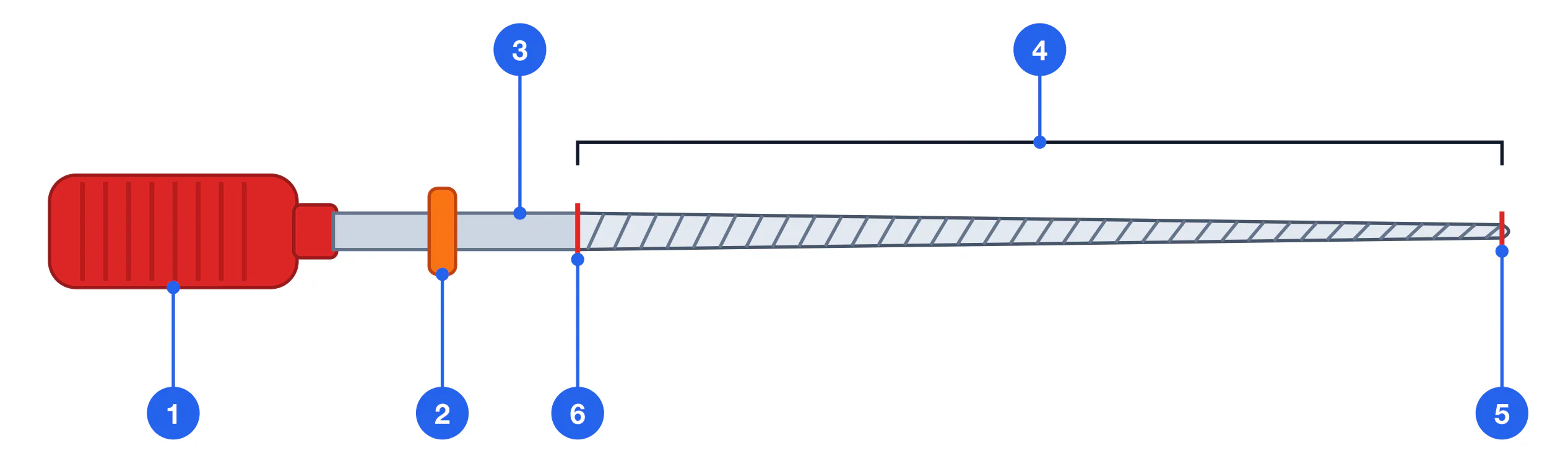
Diameter (D0): 0.06 – 0.80 mm — ISO sizes #06 – #80
Steel file taper: .02 (standard) — 2% — diameter grows 0.02 mm/mm
NiTi taper options: .04 / .06 / .08 — nickel-titanium systems
Instrument length: 21 / 25 / 31 mm — three ISO standard lengths
| Ring color | ISO size | D0 (mm) |
|---|---|---|
| Pink | #06 | 0.06 |
| Grey | #08 | 0.08 |
| Purple | #10 | 0.10 |
| White | #15 | 0.15 |
| Yellow | #20 | 0.20 |
| Red | #25 | 0.25 |
| Blue | #30 | 0.30 |
| Green | #35 | 0.35 |
| Black | #40 | 0.40 |
| White | #45 | 0.45 |
| Yellow | #50 | 0.50 |
| Red | #55 | 0.55 |
| Blue | #60 | 0.60 |
| Green | #70 | 0.70 |
| Black | #80 | 0.80 |
Hand Files: Types, Cross-sections, Indications
K-file K-file — Square or triangular cross-section. Helix angle ≈ 29°. Used by "rotation + pressure" technique: introduce with apical pressure + 1/4-turn rotation + withdrawal. Ideal for negotiating narrow, calcified canals. Manufactured by twisting a steel blank.
H-file Hedstrom file (H-file) — S-shaped cross-section produced by machining (not twisting). Cuts ONLY on the pull stroke — NEVER rotate in the canal or it will fracture. Highly effective for debridement when withdrawing.
K-reamer K-reamer — Rhomboid cross-section, helix angle ≈ 40° (steeper than K-file). More tapered than K-file. Works primarily by rotation — originally designed for reaming. Used in straight or moderately curved canals.
Nickel-Titanium Rotary Systems
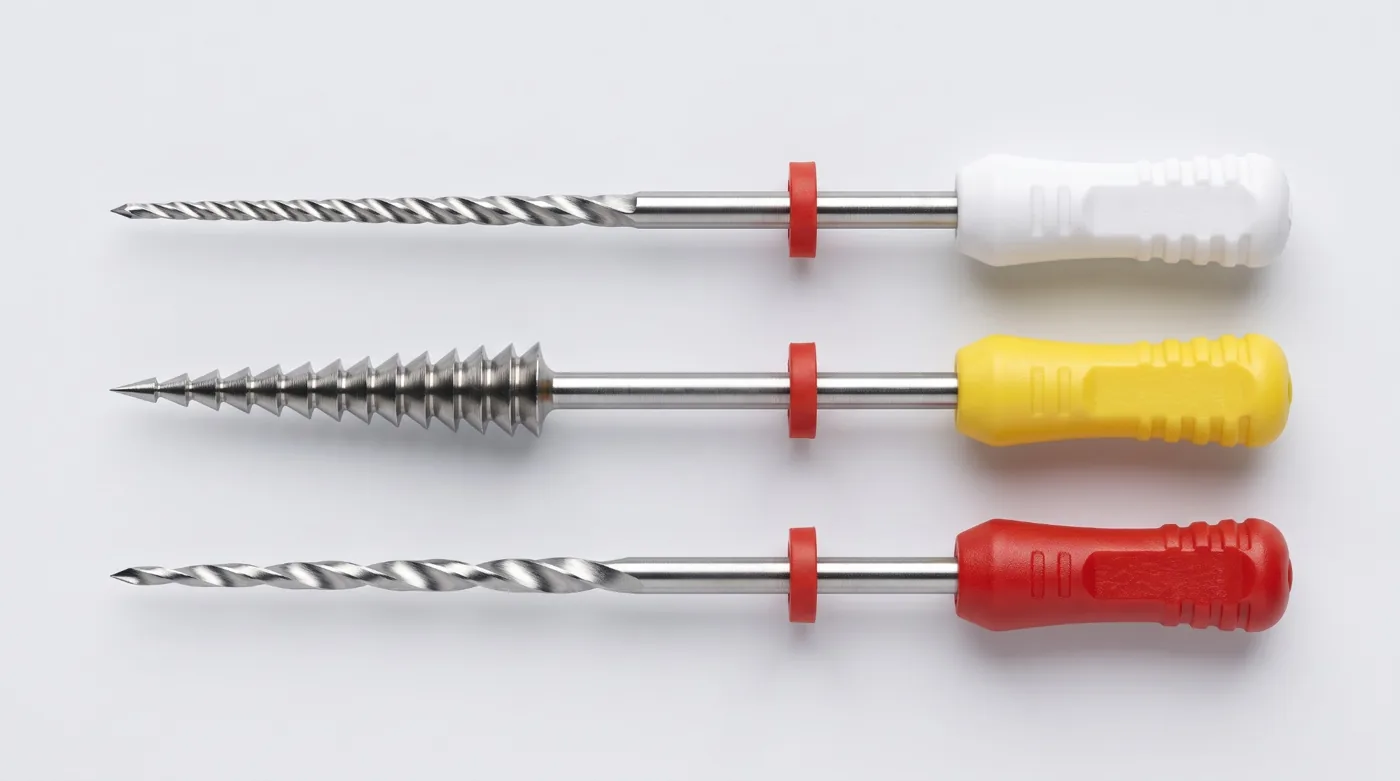
NiTi files are made from an alloy of approximately 55% nickel and 45% titanium. This alloy exhibits superelasticity: the file can flex up to 90° without permanent deformation, unlike steel files which deform at far smaller angles.
Superelasticity arises from a martensitic phase transition within the alloy under stress. However, fatigue fracture of NiTi instruments occurs WITHOUT visible deformation — the instrument fails suddenly when the cyclic fatigue limit is reached. This is the principal hazard of rotary NiTi systems.
ProTaper Gold ProTaper Gold (Dentsply Sirona) — Progressive taper: S1 (.02–.11), S2 (.04–.11), F1 (.07), F2 (.08), F3 (.09). Sequence: shapers S1→S2 open the coronal aspect; finishers F1→F3 complete apical shaping. Gold-wire alloy (Gold heat treatment) provides increased flexibility versus M-wire. One of the most widely used rotary sequences.
WaveOne/Reciproc WaveOne Gold / Reciproc Blue — Reciprocating motion: large clockwise angle + smaller counterclockwise angle. One instrument replaces the full sequence. WaveOne Gold Primary (size 25, .07 taper) covers most canals. Reduces torsional stress and fracture risk.
HyFlex CM HyFlex CM (Coltene) — CM-wire (Controlled Memory): autoclaving partially restores instrument shape, allowing continued use. Deformation is visible before fracture — discard any visibly distorted file immediately.
ProFile/K3 ProFile / K3 (fixed taper) — ProFile: U-shaped cross-section, .04 or .06 taper. Works with crown-down technique. K3 — triangular cross-section with prominent cutting flutes. Both systems are conservative in dentin removal.
NiTi composition: 55% Ni + 45% Ti — nitinol alloy
Flexibility vs steel: 2–3× greater — superelastic NiTi
Fatigue fracture: No warning! — fractures without visible deformation
File replacement: 1–3 uses — NiTi rotary — especially in curved canals
Irrigation: Agents, Sequence, Activation
Irrigation is an integral component of chemo-mechanical canal preparation. Mechanical instrumentation removes only 50–70% of infected dentin; chemical disinfection by irrigants accounts for the remainder.
NaOCl Sodium hypochlorite (NaOCl) 2.5–5.25% — Primary irrigant. Dissolves organic tissue (necrotic pulp remnants) and provides broad-spectrum antimicrobial activity. Do NOT mix with chlorhexidine — precipitate of parachloroaniline (PCA) forms, potentially toxic. 5.25% is more active but more cytotoxic; 2.5–3% is standard.
EDTA EDTA 17% (ethylenediaminetetraacetic acid) — Chelating agent: binds calcium ions → demineralizes peritubular dentin → removes the smear layer. The smear layer is a layer of dentinal debris plugging the tubules. EDTA is applied AFTER NaOCl: NaOCl destroys the organic component of the smear layer; EDTA then removes the inorganic residue.
CHX Chlorhexidine 2% — Substantivity: persists on dentinal walls for up to 12 weeks. Do NOT use immediately after NaOCl — flush with distilled water between them. Used as a final rinse alternative or in patients allergic to NaOCl.
NaOCl 2.5–5.25% — Main irrigant throughout mechanical preparation. Irrigate after each file. Volume: minimum 2 mL per file, total ≥20 mL per canal. (5–10 mL syringe with side-vent needle) (Entire preparation phase)
EDTA 17% — Final stage: introduce 17% EDTA, hold 1 minute. Softens dentin and removes smear layer — opens dentinal tubules for sealer penetration. (17% EDTA gel or solution) (1 minute)
Distilled water flush — Rinse to remove EDTA residues and reaction products. Prevents EDTA-NaOCl interaction (which reduces NaOCl activity). (Sterile distilled water) (30 seconds)
Final NaOCl — Final antibacterial rinse to restore disinfection level after EDTA. (NaOCl 2.5%) (1 minute)
Irrigant activation (PUI) — Passive Ultrasonic Irrigation (PUI): ultrasonic tip vibrates freely within the irrigant, generating acoustic streaming. Alternative: EndoActivator — mechanical activator without heat. Significantly improves irrigant penetration into the apical third and lateral ramifications. (Ultrasonic tip or EndoActivator) (30–60 sec per canal)
Working Length Determination
Working length (WL) is the distance from a coronal reference point (cusp tip, incisal edge) to the apical constriction (minor foramen). The apical constriction lies 0.5–1.0 mm short of the radiographic apex.
Instrumentation and obturation to the major foramen (true apical foramen) is incorrect — it extrudes infection and irrigants into periapical tissues. The target is always the minor foramen.
Radiographic method — Preoperative radiograph with file in canal: measure apparent canal length, subtract parallax correction. Method error: ±0.5 mm. Estimated WL = radiographic length − 1.0 mm. (Periapical radiograph, marker file) (Single measurement)
Electronic apex locator — Root ZX and Raypex 5 are frequency-dependent apex locators of the 3rd and later generations, working at two or more frequencies (Root ZX: 0.4 and 8 kHz). Device measures impedance between file tip and oral mucosa. Accuracy ±0.5 mm. Most precise clinical method. (Apex locator (Root ZX / Raypex 5), file, lip electrode) (Seconds per canal)
WL determination — WL = radiographic length − 0.5–1.0 mm. Confirm with a master-file radiograph. CBCT used for complex anatomy, dilacerations or calcifications. (Radiograph + apex locator combination) (After each significant file change)
Errors in Canal Preparation
Ledge formation — Step-like deformation in the curved portion of the canal from excessive apical pressure. Results in loss of working length. Prevention: pre-curving files, gentle technique, correct sequencing.
Apical transportation (Zipping) — Zip-shaped deformation of the apical portion from over-instrumentation with a straight file in a curved canal. Destroys the apical constriction anatomy. Prevention: crown-down technique, pre-curved files.
Perforation — Instrument penetration through the root wall. Types: furcal, lateral (mid-root), apical. Diagnosis: bleeding in canal, loss of working length, pain on irrigation.
Instrument fracture — Instrument breakage in the canal: fatigue fracture (NiTi rotary — no visible deformation) or torsional fracture (hand files — excessive rotation). Risk factors: >3 uses, curved canal, forced progression. Replace instruments after 1–3 uses in curved canals.
Apical blockage (dentin plug) — Compaction of dentin debris in the apical third forms a "plug" that prevents instruments from reaching the apex. Cause: insufficient irrigation, no recapitulation. Prevention: irrigate after every file, recapitulate with a small file (#10).
Canal Preparation Techniques
Step-back Step-back (apical-to-coronal) — WL is determined first; the apical third is shaped with the MAF (master apical file). Subsequent files are introduced 1 mm shorter than the previous, producing a tapered apical form. Risk: infection can be pushed through the apex on initial file introduction.
Crown-down Crown-down (coronal-to-apical) — Begins with a large file at the canal orifice, progressing to smaller files deeper toward the apex. Advantages: eliminates coronal obstructions before apical shaping; reduces apical extrusion of infected debris; optimal for NiTi rotary systems. Disadvantage: longer; requires precise sequencing.
Balanced forces Balanced forces technique — Hand instrumentation technique (Roane, 1985): the file is inserted with a clockwise rotation of about 90° (engaging dentin), then rotated counterclockwise 120–360° with apical pressure (cutting dentin); after several cycles, a clockwise rotation loads the debris and the file is withdrawn. Maintains canal centring in curved canals better than simple push-pull filing. Technically demanding.
Key terms
Instruments: K-file, H-file, K-reamer, ProTaper Gold, WaveOne Gold, HyFlex CM
Irrigants: NaOCl 2.5–5.25%, EDTA 17%, CHX 2%, PUI
Techniques: Step-back, Crown-down, Balanced forces, Recapitulation
Complications: Ledge, Zipping, Perforation, Instrument fracture