Diagnosis, MTA treatment, fragment retrieval, apical transportation, prevention
ICD-10: —
Root Canal Perforations
A perforation is a pathological or iatrogenic communication between the root canal system and the external root surface or the periradicular tissues (PDL/bone). It is a serious complication: the longer treatment is delayed, the greater the bacterial contamination of the perforation defect and the worse the prognosis.
Furcal Furcal perforation — Location: furcation area. Cause: excessive widening of canal orifices with burs, or over-preparation of the pulp chamber floor. Worst prognosis because of direct communication with the furcal periodontium.
Lateral Lateral (mid-root) perforation — Location: middle third of root. Cause: deviation from the canal axis, excessive file pressure. Prognosis depends on level (more coronal = better) and time to treatment.
Apical Apical perforation — Instrument or irrigant exits through the apical foramen. Often a consequence of apical transportation. Prognosis: moderate with prompt MTA treatment.
Strip Strip perforation — Perforation of the distal wall of the mesial roots of mandibular molars (the so-called "danger zone"): excessive rotary instrumentation thins the dentin on the inner aspect of the canal curvature until penetration into the furcation occurs.
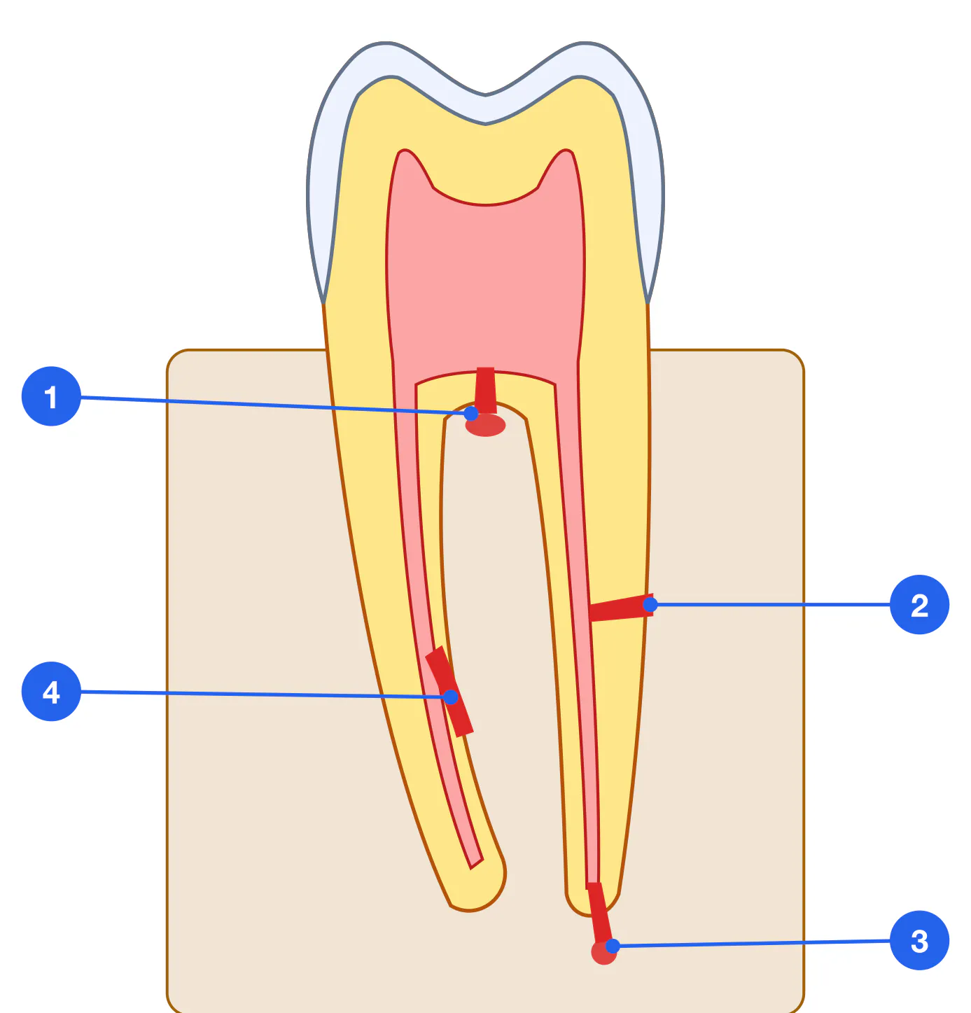
Bleeding in the canal — Sudden appearance of blood on a file in the canal indicates perforation into the periodontal space. Stop immediately and reassess working length.
Loss of working length — The file deflects laterally or loses apical resistance — suspect strip perforation or transportation.
Pain on irrigation — Patient reports sharp pain when NaOCl is irrigated — irrigant is entering periapical/periodontal tissues through the perforation.
Treatment of Perforations: MTA and Alternatives
MTA (Mineral Trioxide Aggregate) is the bioactive cement of choice for perforation repair. It sets in the presence of moisture, generates hydroxyapatite, is highly biocompatible, has a pH of 12 (antibacterial) and is radiopaque.
Isolation and access — Rubber dam is mandatory. Assess perforation location (radiograph, apex locator). Spatial orientation preferably under dental operating microscope (DOM). (Rubber dam, DOM (microscope)) (5–10 min)
Debridement and decontamination — Gentle NaOCl irrigation. If granulation tissue present — careful curettage followed by irrigation. Avoid aggressive removal — tissue destruction worsens prognosis. (NaOCl, curettes) (5 min)
MTA placement — Prepare high-viscosity MTA (Proroot MTA, MTA Angelus). Condense into perforation defect in small increments with MTA carrier or plugger. Confirm tight fill. Moist cotton pellet over MTA for 4 hours. (Proroot MTA / MTA Angelus, MTA carrier, plugger) (5–10 min)
Radiographic verification — Immediate postoperative radiograph. MTA sets in 2–4 hours. Continue endodontic treatment at the next appointment. (X-ray unit) (1–2 min)
MTA MTA (Proroot MTA, MTA Angelus) — Gold standard. Sets in 2–4 hours. Grey MTA may cause coronal discoloration — use white MTA for anterior teeth.
Biodentine Biodentine (Septodont) — Calcium silicate cement. Advantages: fast set (12 min), lower discoloration risk, good biocompatibility. Alternative to MTA when staining is a concern. Slightly lower antibacterial activity.
Super EBA Super EBA — Modified ZOE cement with ortho-ethoxybenzoic acid. Less preferred than MTA/Biodentine but available. Primarily used in apicectomy (retrograde obturation).
Strip Perforation
Strip perforation arises from over-rotary instrumentation in a curved canal, where the inner wall (zone of minimum dentin thickness at the inner curvature) is perforated. Characteristic of the distal walls of the mesial roots of mandibular molars.
Krueger test — When the file is advanced to working length, deviation from the canal axis is felt — strip perforation is suspected. The apex locator shows a perforation signal while the file is in the correct canal.
Bleeding on lateral pressure — Blood appears only when the instrument is pressed against a particular canal wall — the defect is located laterally.
Instrument Fracture in the Canal
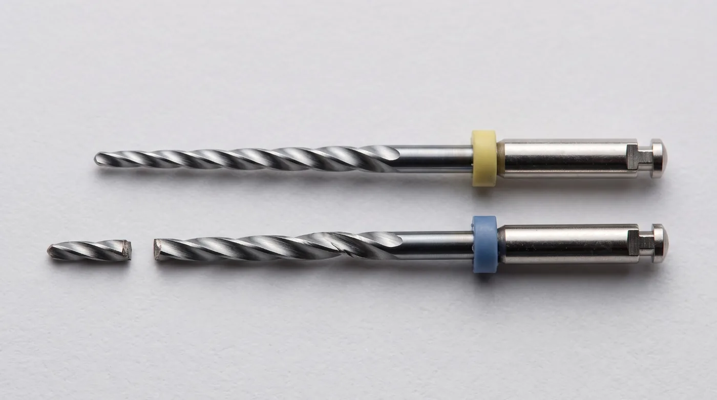
Fracture of an endodontic instrument is one of the most stressful complications for the clinician. Incidence: 1–3% of all instrumented canals (rotary NiTi). Prognosis is determined by several factors, and a fractured instrument does not by itself mean an unfavourable outcome.
Fracture incidence: 1–3% — of rotary NiTi in clinical use
Favourable prognosis: ~80% — short of the apex, no periapical lesion — success without retrieval
Unfavourable prognosis: ~50% — beyond the apex, periapical lesion — lower success
Fragment length: Shorter is better — <5 mm — easier to bypass
Fatigue Cyclic fatigue fracture (NiTi rotary) — Cyclic metal fatigue from repeated flexion in a curved canal. Fractures without visible deformation. Risk factors: curvature >30°, >3 uses, high rotation speed.
Torsion Torsional fracture (hand files) — Excessive twisting: instrument locks in canal while rotation continues. Characteristic of hand files under forceful use. Prevention: light touch, no apical pressure.
Retrieval of Fractured Instruments
Assessment (CBCT) — CBCT diagnosis: precise localization of fragment (distance from orifice, from apex, curvature above fragment). Decision: retrieval attempt vs bypass vs observe. (CBCT unit) (5 min (image analysis))
Ultrasonic retrieval — Standard approach when microscope is available. Specialised ultrasonic tips (Endoextractor, IRS, ProUltra EMS) generate vibration at the proximal end of the fragment → loosen the "wedge" → fragment rotates counterclockwise and exits. Prerequisites: fragment in a straight segment, visible under DOM. (Ultrasonic scaler, tips, DOM) (10–30 min)
Masserann tubes — Hollow tubular extractor system. Tube introduced over fragment; rotation engages and removes it. Requires wider canal preparation — suitable only for coronally positioned fragments. (Masserann kit) (15–20 min)
Bypass attempt — If retrieval fails: attempt to bypass with a small file (#06–#08). Goal: establish patency alongside fragment → determine WL → continue preparation. (K-file #06, #08, #10, EDTA cream) (20–40 min)
Leave and monitor — If fragment is in the apical curved portion, no periapical pathology, and retrieval fails — obturate to the fragment level and monitor. Without periapical pathology prognosis is comparable to successful treatment. (Dynamic recall: 3–6–12 months)
| Factor | Favourable | Unfavourable |
|---|---|---|
| Fragment position | Coronal third, straight segment | Apical third, curved canal |
| Periapical pathology | None | Periapical lesion present |
| Pre-fracture infection | Vital pulp, no infection | Pulp necrosis, chronic inflammation |
| Fragment length | Short (<5 mm) | Long (>5 mm) |
| Visibility under DOM | Fully visible | Not visible (too deep) |
Prevention of Instrument Fracture and Apical Transportation
Visual inspection — Inspect instrument before and after each use: flute deformation, unwinding, scratches. NiTi rotary files show no deformation before fracture — replace strictly per protocol. (Before each use)
File replacement protocol — Hand K-files: 5–7 uses. Rotary NiTi in straight canals: up to 3–5 uses. Rotary NiTi in curved canals (>25°): 1–2 uses. Single-use systems (WaveOne, Reciproc): 1 patient. (Per use tracking protocol)
Pre-curving files — Before introducing into a curved canal — pre-curve (bend the file with fingers to match canal curvature). Especially important for K-files on initial negotiation of tight curved canals. (Before introduction into each curved canal)
Crown-down and torque control — Crown-down technique reduces apical stress. Use NiTi rotary with a torque-limited motor — auto-reverse on torque threshold prevents torsional fracture. (Torque-limited endodontic motor) (During all rotary instrumentation)
EDTA cream lubrication — RC Prep, Glyde, or EDTA cream introduced ahead of the file. Lubricates canal walls, reduces friction → lower torque → reduced fracture risk. (RC Prep / Glyde) (Before each file)
Key terms
Complications: Perforation, Strip perforation, Apical transportation, Instrument fracture
Treatment: MTA, Biodentine, Ultrasonic retrieval, Masserann, Bypass
Prevention: Crown-down, Pre-curving, EDTA cream, Torque-control motor