Biomechanics, Applegate's rules, clasp systems, Ante's law and the IROPZ index
ICD-10: K08.1
Definition and Clinical Significance
The Kennedy Classification (1925) is the most widely accepted topographic system for describing partially edentulous arches. Proposed by Edward Kennedy in the USA; in 1954 Applegate added 8 rules for application. It divides all defects into 4 classes based on the relationship of edentulous spaces to the remaining teeth.
Clinical significance: the classification directly determines the biomechanics of the planned prosthesis (support type — tooth-borne, tissue-borne or tooth-tissue-borne), clasp position, direction of prosthesis displacement under load, and long-term prognosis for abutment teeth.
Author: Edward Kennedy — 1925, USA
Classes: 4 main classes — + subclasses by number of additional defects
Applegate rules: 8 rules — Refine classification application
First molar PDL area: 433 mm² — Periodontal ligament (Ante's law)
Kennedy Classes
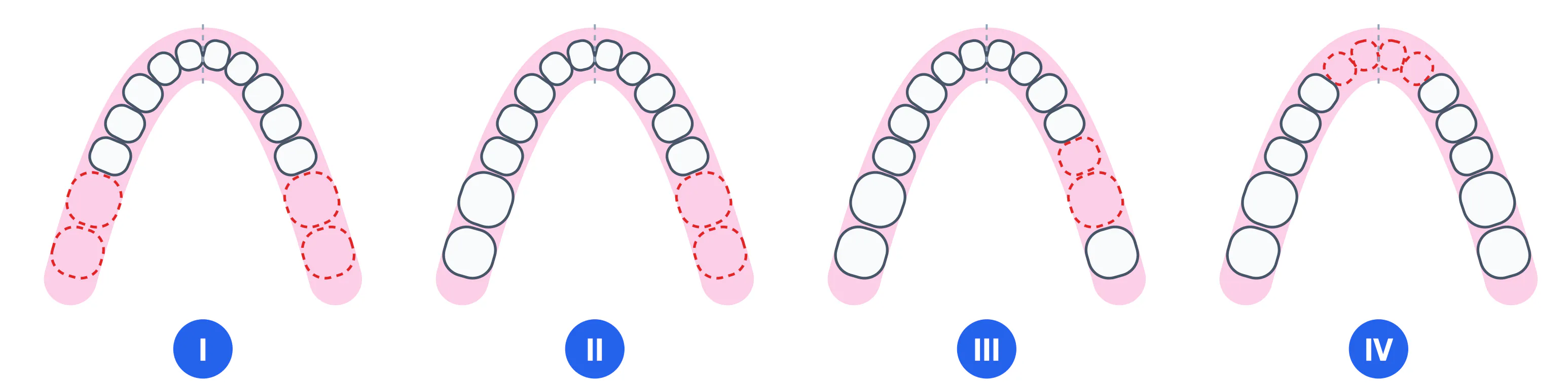
Class I Bilateral free-end defect — Missing teeth on both sides posterior to the last abutment teeth. Prosthesis has combined tooth-tissue-borne support. Class 1 lever: fulcrum = most distal clasp; masticatory force on extension bases creates torque on the abutment tooth.
Class II Unilateral free-end defect — Missing teeth on one side posterior to the last abutment (free-end unilateral). Asymmetric prosthesis. Tendency to rotate about the axis of the contralateral clasp.
Class III Bounded posterior defect — Defect has abutment teeth both anterior and posterior. Purely tooth-borne support — prosthesis does not load the mucosa under function. Fixed bridge possible for short spans.
Class IV Bounded anterior defect — A single defect crossing the midline, bounded by posterior teeth. No subclasses. If there are additional edentulous areas posteriorly, the most posterior area determines the class and the anterior space counts as a modification — the arch is then no longer Class IV. Aesthetic restoration required — RPD or implants.
Biomechanics of the RPD
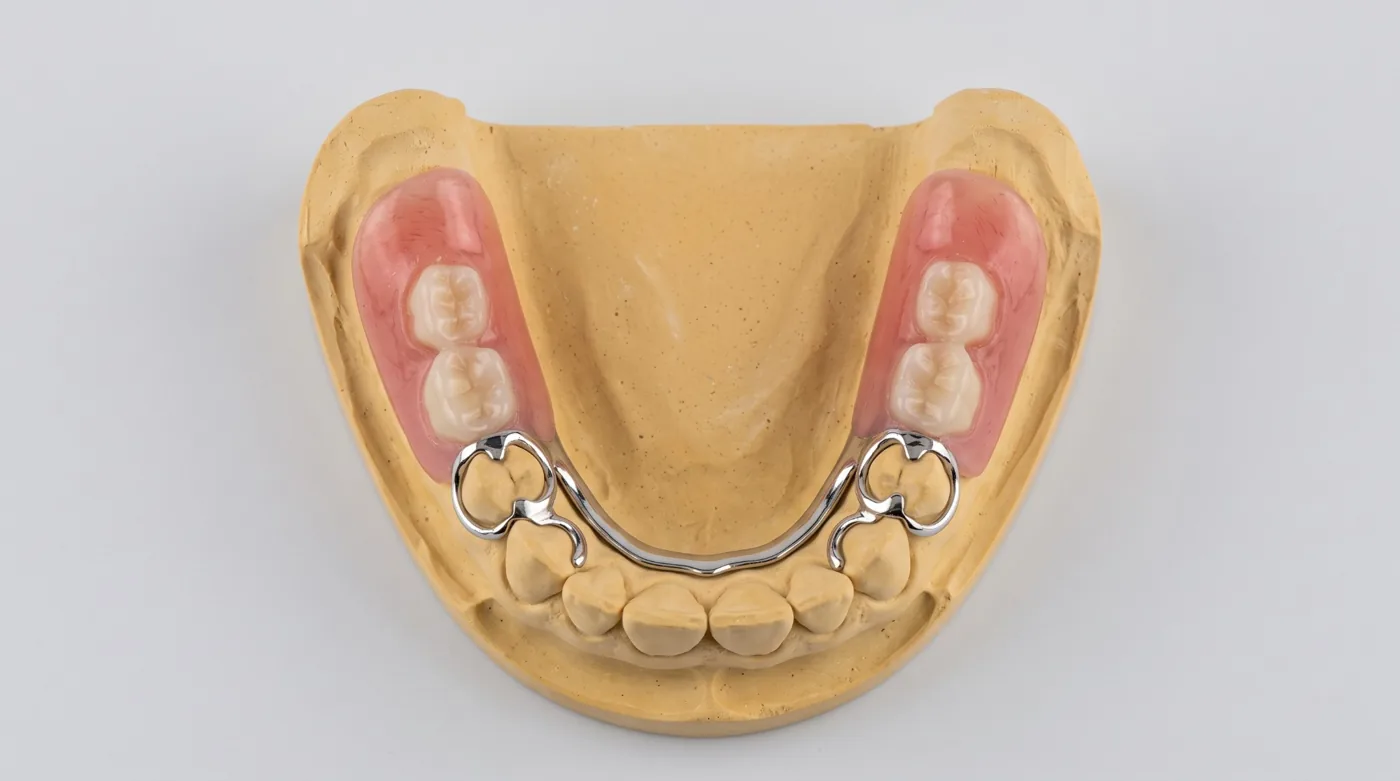
Understanding RPD biomechanics is essential to prevent abutment overload. For free-end extensions (Classes I and II), the RPD acts as a Class 1 lever: the fulcrum is the most distally placed clasp assembly. During mastication, the extension base is loaded apically (mandible) or occlusally (maxilla), creating a tipping moment transmitted to the abutment tooth via the clasp.
The free-end saddle is tooth-tissue-borne: load is transmitted both through the clasp to the abutment tooth (via the periodontal ligament) and through the denture base to the residual ridge mucosa and underlying bone. In contrast, a bounded defect (Class III) is purely tooth-borne — load passes through clasps on both abutments without mucosal contact.
Classes I & II: Combined support — Tooth-tissue-borne
Classes III & IV: Tooth-borne only — No load on mucosa
Class 1 lever: Fulcrum = distal clasp — Tipping moment on abutment
Abutment protection: RPI / RPA clasp — Minimal tooth contact under load
Ante's Law
Ante's law (1926) states: the total periodontal ligament (PDL) surface area of all abutment teeth must equal or exceed the total PDL surface area of all teeth to be replaced. The law is applied in fixed bridge and clasp RPD planning to assess the permissibility of loading abutment teeth.
Calculation example: missing tooth 36 (433 mm²) + 37 (431 mm²) = 864 mm² of PDL to replace. Proposed abutments — 35 (215 mm²) + 38 (431 mm²) = 646 mm². Ante's law is NOT met → fixed bridge is contraindicated; RPD or implants are indicated.
| Tooth | PDL area (mm²) | Planning significance |
|---|---|---|
| Central incisor | 204 mm² | Weak abutment for long-span bridges |
| Lateral incisor | 179 mm² | Smallest PDL area — caution under occlusal load |
| Canine | 273 mm² | Best anterior abutment tooth (long root) |
| First premolar | 220 mm² | Good abutment with healthy periodontium |
| Second premolar | 215 mm² | Comparable to first premolar |
| First molar | 433 mm² | Best posterior abutment tooth |
| Second molar | 431 mm² | Virtually equal to first molar |
Prosthesis Selection by Kennedy Class
| Kennedy Class | Optimal Prosthesis | Condition | Alternative |
|---|---|---|---|
| Class I | RPD (clasp or attachment-retained) | Adequate abutment teeth present | Complete denture if no abutments remain |
| Class II | RPD (unilateral distal extension) | Support anterior to defect | Fixed bridge for short spans |
| Class III | Fixed bridge | Short span (1–2 teeth), healthy abutments, Ante's law met | RPD for long spans or compromised abutments |
| Class IV | Partial RPD / Implants | Aesthetics + phonetics | Fixed bridge for 1–2 missing teeth |
All 8 of Applegate's Rules
Rule 1 — Classify after extractions — The arch must be classified AFTER all planned extractions have been performed, not before.
Rule 2 — Third molars absent — If third molars are absent and are not to be replaced, they should not be used as class-defining posterior boundaries.
Rule 3 — Most posterior defect determines the class — When multiple defects exist, the class is determined by the most posteriorly located defect.
Rule 4 — Subclasses from additional defects — Additional edentulous areas (other than the primary class-defining defect) determine the subclass number.
Rule 5 — Class IV overrides other classes — If a Class IV situation exists (anterior bounded defect crossing the midline), it takes precedence in classification regardless of other defects present — and receives no subclass.
Rule 6 — Anterior teeth missing with Class III — If teeth are missing anterior to the Class III-defining defect, they do not change the class — they become subclasses.
Rule 7 — One additional defect per subclass — Each separate additional edentulous area increases the subclass number by one. Two additional defects = subclass 2.
Rule 8 — Class III subclass 1 = one additional defect — Class III subclass 1 indicates exactly one additional bounded edentulous area beyond the primary posterior bounded defect.
Clasp Systems for RPDs
Clasp system selection determines RPD biomechanics, abutment tooth loading and aesthetics. Three principal systems are used in contemporary prosthodontics.
RPI system Rest – Proximal Plate – I-bar — Most physiological clasp system for free-end extensions. Mesial occlusal rest, proximal plate guiding the prosthesis, I-bar retainer engaging tissue undercut. Advantages: minimal tooth contact, no buccal lever arm on the abutment, good aesthetics. Indicated for Class I and II RPDs.
RPA system Rest – Proximal Plate – Akers clasp — Used when favourable tissue undercut is absent (I-bar cannot engage). Akers buccal arm encircles the abutment from the buccal aspect. Slightly greater torque on the abutment compared with RPI.
Circlet / back-action clasp Ring clasp for tilted molars — Used on molars that have tipped towards the edentulous space. The arm encircles nearly 360°, engaging retention from the distolingual aspect. Indicated for distally tilted last molar abutments.
IROPZ Index (Milikevich)
IROPZ (Russian ИРОПЗ — index of destruction of the occlusal surface of a tooth) was developed by V.Yu. Milikevich; it has no established equivalent in English-language literature, so the original abbreviation is used. It is calculated as the ratio of the defect area (cavity or destroyed surface) to the total occlusal surface area of the tooth: IROPZ = defect area / occlusal surface area.
The index ranges from 0.0 to 1.0. Practical rule: the higher the IROPZ, the more invasive the restoration required to rebuild the tooth.
IROPZ ≤ 0.3 Inlay / onlay — Small defect — indirect ceramic or metal inlay is indicated. Maximum tooth tissue preservation.
IROPZ 0.3–0.6 Inlay or crown — Borderline zone. Decision is made based on tooth location and aesthetic demands. Preference for inlay.
IROPZ 0.6–0.8 Crown on core — Significant destruction — a cast post-core build-up or composite core build-up with subsequent full-coverage crown is required.
IROPZ > 0.8 Post-retained restoration — Crown almost entirely destroyed to the gingival level. Cast post-core crown or post-retained crown required. If crown-to-root ratio is unfavourable, consider extraction and implant.