Wear mechanisms, BEWE index, VDO loss pathogenesis, Michigan splint and the Dahl concept
ICD-10: K03.0
Definition and Epidemiology
Pathological tooth wear is the progressive loss of hard tooth tissue exceeding physiological norms, resulting from mechanical, chemical or combined factors. Differentiating the underlying mechanism is critical: attrition, erosion, abrasion and abfraction present with distinct wear patterns, different aetiologies and require different management strategies.
Affects 12–15% of the population, most commonly in men aged 35–50. Without treatment, loss of vertical dimension of occlusion (VDO) leads to TMJ dysfunction, periodontal overload, neuromuscular fatigue and aesthetic complaints.
ICD-10: K03.0 — Excessive attrition of teeth
Prevalence: 12–15% — Population, commoner in men 35–50
BEWE index: 0–3 per sextant — Erosive wear screening (Bartlett, Ganss, Lussi, 2008)
Degrees (Bracco): 0–III — By area and depth of destruction
Differentiating Wear Mechanisms
Accurate diagnosis of the wear mechanism guides etiological treatment. Four principal mechanisms have characteristic clinical presentations that should be identified at an early stage.
Attrition Tooth-to-tooth contact (attrition) — Smooth, polished, matching wear facets on antagonist teeth. Location: occlusal surfaces and incisal edges. Principal cause: bruxism (sleep or awake). Hallmark: mirror-image facets on upper and lower teeth that only occlude during parafunctional contact.
Erosion Chemical dissolution (erosion) — Smooth concave surfaces without corresponding antagonist facets. In GERD: palatal surfaces of upper anteriors (gastric acid bathes the palate). In bulimia nervosa: same location. In dietary acids: buccal surfaces. Key feature: no facet match between upper and lower teeth.
Abrasion Abrasive wear (abrasion) — V-shaped or wedge-shaped notches at the cervical margin. Cause: horizontal toothbrushing with abrasive toothpaste. Surface is matt (not polished). Location: cervical third of crown on buccal aspect.
Abfraction Stress-fatigue lesions (abfraction) — Wedge-shaped cervical lesions from stress concentration at the cervical region caused by lateral occlusal forces. Surface is sharp and angular. Theory: eccentric loads create tensile stress zones at the cervix → enamel microcracking. Often combines with erosion and abrasion.
Degrees by Bracco
Degree 0 Physiological — Wear within enamel of incisal edges and cusps. No treatment required.
Degree I Mild — Enamel wear on occlusal surfaces and incisal edges. Dentin not exposed or barely visible. Crown height minimally reduced. VDO unchanged.
Degree II Moderate — Enamel and dentin wear up to 2/3 of crown height. Dentin exposed, possible hypersensitivity. VDO reduced 2–3 mm. Neuromuscular adaptation present.
Degree III Severe — Wear to the gingival level or cervix. Crown virtually destroyed. Marked VDO loss (>4 mm), TMJ dysfunction, pain, paresthesia.
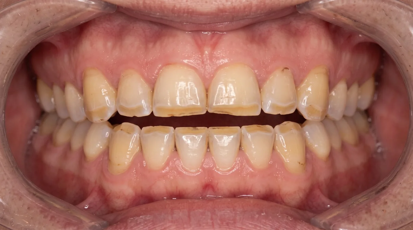
Degrees by Grozovsky
A. L. Grozovsky's classification grades the depth of hard-tissue loss by crown height. Unlike the Bracco scale, which describes the clinical picture as a whole (dentin, VDO, TMJ), it considers only how much of the crown height has been lost.
Degree I Up to 1/3 of crown height — Loss of enamel and some dentin within the occlusal (incisal) third of the crown.
Degree II 1/3 to 2/3 of crown height — Loss of enamel and dentin up to 2/3 of crown height; dentin exposed over a large area.
Degree III More than 2/3 of crown height — Loss of more than 2/3 of crown height, down to the gingival level.
BEWE Index (Basic Erosive Wear Examination)
BEWE is a standardized index for screening erosive tooth wear, proposed by Bartlett, Ganss and Lussi (2008); it is not a WHO recommendation. The highest score in each of the 6 sextants is recorded and then summed.
| BEWE score | Criterion | Clinical appearance |
|---|---|---|
| 0 | No erosion | Surface intact |
| 1 | Initial loss | Surface texture change, loss of lustre, no hard tissue loss |
| 2 | Loss < 50% of surface | Visible hard tissue loss, occupies less than half the surface |
| 3 | Loss ≥ 50% of surface | Extensive hard tissue loss, dentin exposure, possible pulp involvement |
BEWE sum 0–2 No measures — Routine monitoring, oral hygiene instruction.
BEWE sum 3–8 Preventive measures — Remineralization therapy, dietary and hygiene counseling, eliminate etiological factors.
BEWE sum 9–13 Prevention + restorations — Monitor every 6–12 months, prevention, restorations on most affected teeth as required.
BEWE sum ≥ 14 Full rehabilitation — Comprehensive prosthodontic treatment: VDO increase, full-coverage crowns or composite restorations on all teeth.
Pathogenesis of VDO Loss
Progressive tooth wear does not inevitably cause proportional VDO loss. The neuromuscular system partially compensates through dento-alveolar adaptation (the Dahl phenomenon): as anterior teeth wear, the posterior teeth undergo apparent eruption, maintaining the lower facial height. This is why the amount of tooth tissue lost often exceeds the actual reduction in VDO.
When compensation is exceeded or decompensation occurs: the masticatory muscles (masseter, temporalis) experience chronic tension → tension-type headaches (TTH), condylar displacement and articular disc compression in the TMJ, and neuromuscular fatigue. Advanced cases show deepening of the nasolabial and labiomental folds and reduction in lower facial height.
Michigan Splint for Bruxism
The Michigan splint (flat-plane stabilization splint) is the gold standard for conservative management of sleep bruxism and TMJ dysfunction. It is a full-arch hard acrylic appliance fabricated for the maxillary arch.
Design requirements — Full coverage of all maxillary teeth. Thickness: 1.5–2 mm. In centric occlusion — even, simultaneous bilateral point contacts of the supporting cusps of all mandibular teeth against the flat splint surface (centric stops). In lateral and protrusive excursions — canine and anterior guidance along the splint ramp, which disoccludes the posterior teeth. (Hard PMMA acrylic — laboratory fabricated)
Wear regimen — Nocturnal wear 6–8 hours per night. Do not remove during the initial adaptation weeks. First review appointments at 2 weeks and 1 month for occlusal adjustment of the splint. Re-adjust every 3–6 months. (Ongoing, minimum 6 months)
Mechanism of action — Redistributes masticatory load. Unloads the TMJ articular disc. Eliminates harmful parafunctional contacts. Allows masticatory muscle relaxation. May facilitate condylar repositioning in centric relation.
The Dahl Concept
The Dahl concept (Bjørn Dahl, 1975) is a minimally invasive method of creating posterior inter-occlusal space when managing worn anterior teeth. It avoids the need for immediate full-mouth crown rehabilitation by exploiting dento-alveolar compensation.
Technique: direct or indirect composite restorations are placed on the lower anterior teeth (commonly 31, 32, 41, 42), increasing the OVD by 2–3 mm and leaving the posterior teeth out of contact. Over 6–12 months, dento-alveolar compensation occurs: the posterior teeth gradually re-erupt (extrusion) while the anterior teeth intrude slightly, restoring posterior occlusal contacts. The anterior teeth are then restored with definitive restorations.
Full-Mouth Rehabilitation Protocol
Degree I — monitoring and prevention — Remove cause (bruxism → Michigan splint). Fluoride applications, high-fluoride toothpaste (>1450 ppm F). Direct composite restorations for aesthetic concerns. BEWE monitoring every 6 months. (Michigan splint, fluoride varnish, composite)
Degree II — restore VDO — Phase 1: Michigan splint for 3–6 months to stabilize VDO and achieve neuromuscular adaptation. If VDO increase needed: Dahl concept (direct composite) or provisional crowns. Phase 2: definitive full-arch PFM or zirconia crowns. (PFM or zirconia crowns, Michigan splint) (6–12 months)
Degree III — full rehabilitation — Diagnostic wax-up on semi-adjustable articulator → provisional acrylic crowns for 3–6 months → evaluate function, phonetics and aesthetics → definitive PFM or zirconia crowns. Endodontic treatment only where indicated. Cast post-core build-ups for grossly broken-down teeth. Implants and crowns for missing teeth. Permanent Michigan splint for ongoing bruxism. (Cast post-cores, PFM / zirconia crowns, implants) (12–24 months)