Anatomy, DC/TMD 2014 protocol, imaging, Michigan splint, pharmacotherapy and surgery
ICD-10: K07.6
TMJ Anatomy
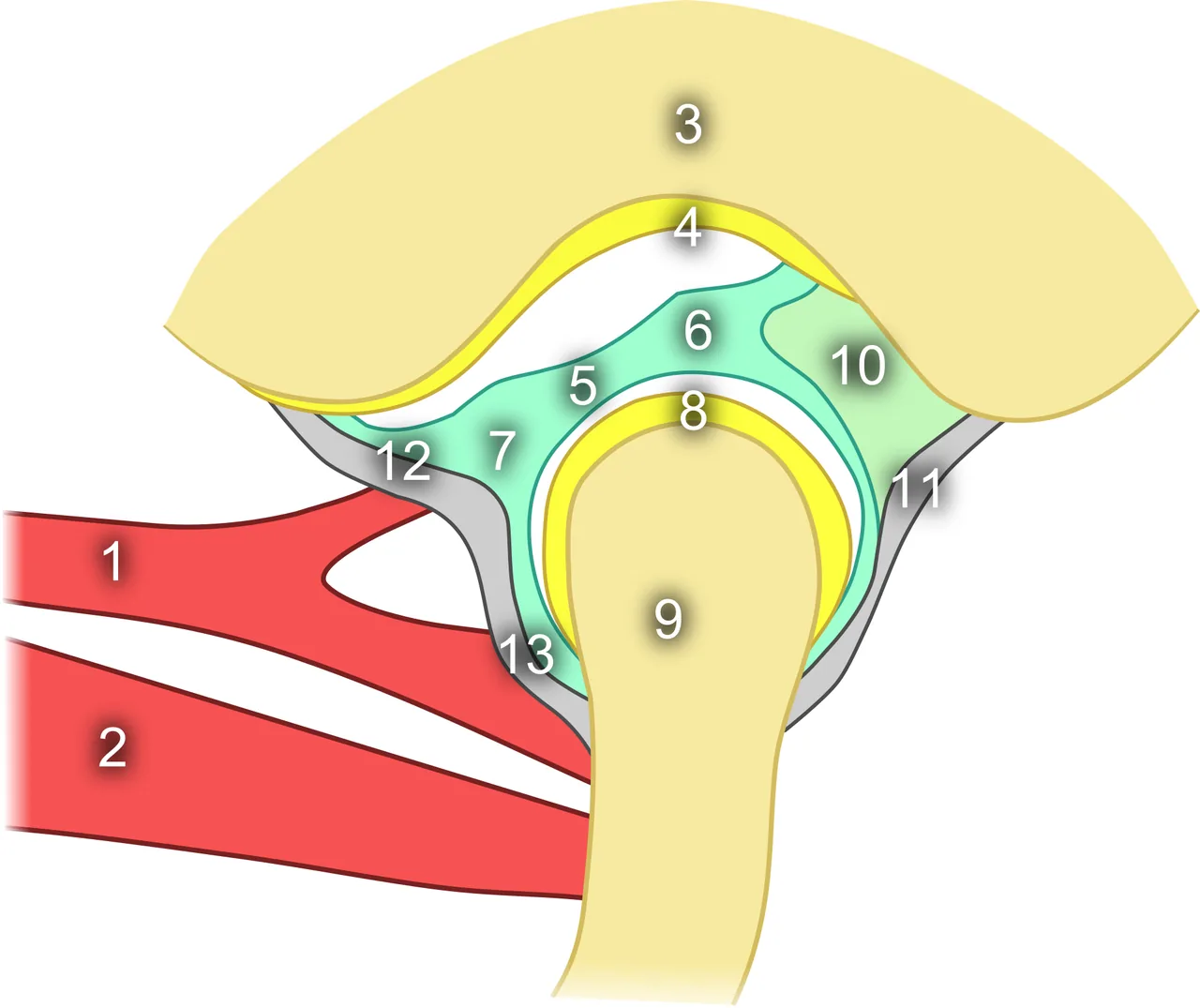
The TMJ is a ginglymoarthrodial (hinge-and-glide) synovial joint with bilateral functional interdependence. The articular disc (discus articularis) is a biconcave fibrocartilage plate that is avascular and aneural in its central (intermediate) zone. The disc divides the joint space into two compartments: the upper (superior) compartment — responsible for translational movements — and the lower (inferior) compartment — responsible for rotational hinge movements.
The posterior disc attachment (bilaminar zone) comprises two layers: the superior stratum (elastic tissue that retracts the disc posteriorly on mouth closing) and the inferior stratum (inelastic collagen fibers). Damage to the bilaminar zone is a key factor in irreversible disc displacement. The superior belly of the lateral pterygoid muscle inserts into the anterior-medial pole of the disc and pulls it forward during mouth opening.
ICD-10: K07.6 — Diseases of TMJ
Diagnostic standard: DC/TMD 2014 — Diagnostic Criteria for TMD (revised)
MRI: Gold standard — For disc position assessment
Prevalence: 20–30% — Symptomatic, F > M (3:1)
DC/TMD Classification (2014)
The Diagnostic Criteria for TMD (DC/TMD, 2014) is the current gold-standard diagnostic framework, superseding RDC/TMD (1992). It uses a two-axis system: Axis I — physical (clinical) diagnoses; Axis II — psychosocial screening using PHQ-4 (PHQ-2 for depression + GAD-2 for anxiety) and the Graded Chronic Pain Scale (GCPS).
Group I Muscle disorders — Myofascial pain with or without limited opening. Palpation tenderness of masticatory muscles, diffuse facial pain, morning stiffness. Diagnostic requirement: familiar pain reproduced by palpation and/or jaw movement.
Group II Disc displacements — IIa — displacement with reduction (click on opening and closing, disappears on protrusive opening provocation). IIb — displacement without reduction, with limitation (closed lock, interincisal opening <40 mm, does not resolve with traction). IIc — displacement without reduction, without limitation (disc permanently displaced, opening normal).
Group III Arthritis and arthrosis — IIIa — arthralgia (pain on movement or joint loading palpation). IIIb — osteoarthritis (crepitus + pain + radiographic changes). IIIc — osteoarthrosis (crepitus, radiographic changes, no pain). CBCT required to confirm bony changes.
Clinical Features
TMJ pain — Dull, aching. Worsened by chewing, wide mouth opening and on waking. Radiates to temple, ear and neck. Characteristic of arthralgia and myofascial pain.
TMJ clicking — Click on opening = disc displacement with reduction (IIa). Reciprocal click (on opening and closing) = IIa. Click that disappears on protrusive opening = differentiating sign for IIa from other causes.
Limited mouth opening — Normal: >40 mm (interincisal distance). 30–40 mm = moderate limitation. <25 mm = severe limitation (closed lock, IIb). Ipsilateral deviation of the mandible towards the locked side on opening.
Crepitus — Coarse grinding sound = sign of osteoarthrosis (IIIb–IIIc). Differs from a click: continuous without a fixed point. Auscultate with a stethoscope at the preauricular point.
Morning masticatory muscle stiffness — Characteristic of muscle disorders (Group I). Patient reports jaw "tightness" on waking. Associated with sleep bruxism or nocturnal hypoxia.
DC/TMD Examination Protocol
Axis II — psychosocial screening — PHQ-4 (4 questions): PHQ-2 — depression, GAD-2 — anxiety. Total score ≥3 = screen-positive; psychology/psychiatry referral indicated. GCPS — intensity and impact of chronic pain on daily function. (PHQ-4 and GCPS questionnaires)
Mouth opening measurements — Pain-free opening (PFO), maximum assisted opening (MAO), left and right lateral excursion, protrusion. Normal opening: 40–55 mm. Record path of opening: straight / S-curve / deviation. (Millimetre ruler (plastic))
Muscle palpation (pain intensity 0–3) — m. masseter (superficial and deep head): 3 sites × 2 sides. m. temporalis (anterior, middle, posterior): 3 sites × 2. m. pterygoideus medialis (intraoral). m. digastricus posterior (submandibular). Record: 0 = no pain, 1 = mild, 2 = moderate, 3 = severe. (RDC/DC-TMD palpation chart)
Imaging — OPG / panoramic radiograph: bony changes (flattening, sclerosis, osteophytes) — but NOT for disc assessment. MRI of the TMJ: gold standard — disc position in closed and open mouth (displacement, deformity, effusion). CBCT: condylar erosion, osteophytes, fractures — bony changes only. (MRI of the TMJ mandatory for IIb–IIc and all surgical planning)
Treatment
Stabilization splint (first line) — Michigan flat-plane splint — full-arch hard acrylic for the maxilla. Thickness 1.5–2 mm. Even bilateral contacts of all supporting cusps in centric occlusion (centric stops). Canine guidance along the splint ramp disoccluding the posterior teeth during all excursive movements. Nocturnal wear 6–8 hours. Mechanism: distributes joint load, masticatory muscle relaxation, possible condylar repositioning towards centric relation. (Michigan stabilization splint) (3–6 months)
Pharmacotherapy — NSAIDs: ibuprofen 400 mg × 3 times/day × 7–10 days (for acute pain). Muscle relaxants: tizanidine 2–4 mg nocte (for severe muscle spasm). Botulinum toxin A: 20–25 units into masseter bilaterally for persistent myofascial pain refractory to splint therapy — effect duration 3–6 months. (Ibuprofen, tizanidine, botulinum toxin A)
Physiotherapy — TMJ exercise programme: stretching and coordination exercises (Rocabado protocol). Moist heat applications. Ultrasound therapy. TENS (transcutaneous electrical nerve stimulation). Normalises muscle tone, improves microcirculation.
Occlusal adjustment (second line) — Only AFTER achieving sustained pain remission on splint therapy (≥3 months). Selective occlusal adjustment at premature contacts. Orthodontic treatment. Prosthodontic rehabilitation. Never initiate during the active pain phase!
Surgical treatment (third line) — Arthrocentesis: lavage of the upper joint compartment under local anaesthesia; indicated for closed lock (IIb) unresponsive to conservative treatment after 4–6 weeks. Arthroscopy — for IIb–IIc, adhesive capsulitis. Open arthroplasty — severe osteoarthrosis, ankylosis, condylar fractures. (Only when conservative treatment has failed)