Requirements for cavity shape, walls, margins and floor. Black's classical principles and the modern minimally invasive approach
ICD-10: —
Overview
Cavity preparation is the mechanical removal of carious tooth tissue to create a cavity form that enables reliable retention and durable function of a restorative material. Preparation quality is one of the primary determinants of restoration longevity: an incorrectly shaped cavity leads to fracture, marginal breakdown, and secondary caries.
G.V. Black (1908) systematized preparation principles developed for the era of amalgam restorations, which require mechanical retention and provide no adhesive bond. Most of Black's principles remain valid today; however, the modern concept of Minimal Intervention Dentistry requires adapting several of them to the properties of adhesive composites.
Preparation steps: 5–6 — by Black: access → extension → necrectomy → shaping → finishing
Wall angulation: 90° — to enamel surface for adhesive restorations
Cavity floor: Flat — or slightly convex — even load distribution
Dentin over pulp: ≥1 mm — in dentinal caries; ≥0.5 mm in deep caries
Preventive extension: Limited — modern principle: only as far as sound tissue
Principle: Min. invasion — preserve maximum healthy tooth structure
Black's Classical Principles
Black described six sequential steps for cavity preparation. This system was designed for amalgam restorations, which require mechanical retention and cannot bond to tooth tissue. Nevertheless, the first four steps apply regardless of material.
1. Cavity access (opening) — Removal of overhanging enamel margins to provide direct access to the affected dentin. Instrument: round bur in high-speed handpiece. Goal: complete visualization of cavity floor and walls. (Step 1)
2. Preventive extension — Extending cavity margins to self-cleansing surfaces not susceptible to caries (immune zones). For amalgam: 'extension for prevention' — extend to self-cleansing areas. For adhesive systems: extend only to the boundary of infected tissue. (Step 2)
3. Necrectomy (caries removal) — Removal of all infected and softened dentin. Instruments: round bur in low-speed handpiece, spoon excavator. Endpoint: firm dentin on probing, no pain on gentle pressure. (Step 3)
4. Cavity shaping (resistance form) — Creating cavity geometry that distributes occlusal forces evenly and prevents restoration fracture. Requirements: flat or slightly convex floor, walls perpendicular to floor (Class I), 90° cavosurface angle. (Step 4)
5. Retention form — For amalgam: box shape with walls slightly convergent towards the occlusal surface (undercut), retention grooves in axio-pulpal line angles. For adhesive systems: mechanical retention is unnecessary — the adhesive bond provides sufficient retention through micromechanical and chemical attachment. (Step 5)
6. Finishing cavity walls and margins — Smoothing walls, removing unsupported and hanging enamel. For adhesive restorations: 90° cavosurface angle (no bevel on Class I and II margins). For cast metal inlays: 45° enamel bevel. For ceramic inlays a bevel is contraindicated — the margin is prepared as a 90° butt joint, otherwise the thin ceramic at the bevel chips. (Step 6)
Requirements for Cavity Elements
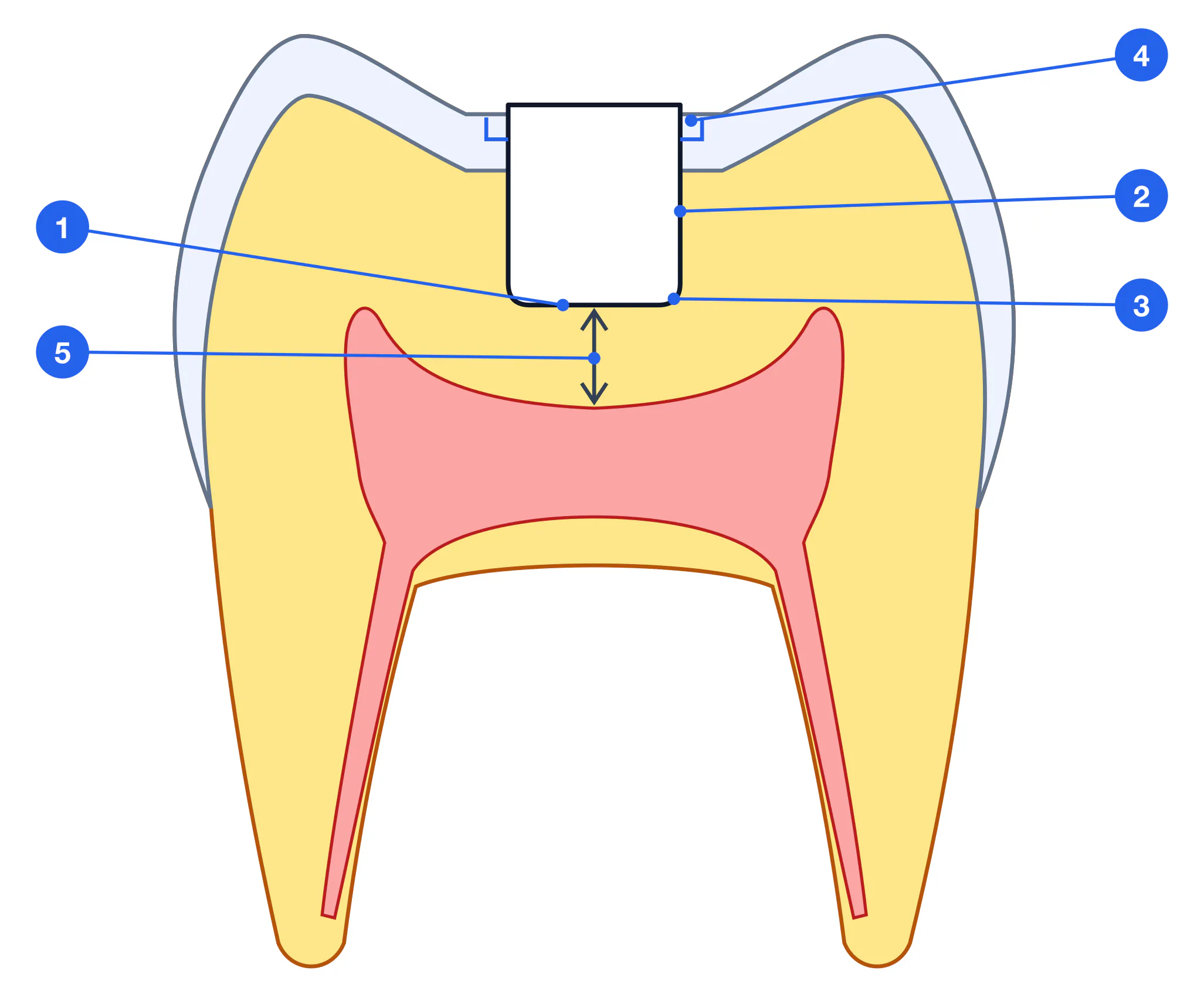
Each element of the cavity — floor, walls, angles and margins — has specific requirements; failure to meet them leads to clinical failure.
| Element | Requirement | Violation → consequence |
|---|---|---|
| Floor | Flat or slightly convex, perpendicular to long axis | Slanted floor → uneven load distribution → fracture |
| Walls | Straight, smooth, perpendicular to floor (Class I–II) | Tapered walls → amalgam fallout; too thin → tooth fracture |
| Internal angles | Rounded (not sharp) | Sharp angles → stress concentration → material crack |
| Cavosurface margins | 90° to enamel surface (for composite) | Bevel on Class I → thinned enamel → stress fractures |
| Depth | Complete removal of infected dentin | Incomplete removal → secondary caries → treatment failure |
| Volume | Minimum necessary | Over-preparation → weakened tooth → fracture |
Modern Minimally Invasive Approach
The concept of Minimal Intervention Dentistry (MID, FDI World Dental Federation, 2002) revisits Black's classical principles for adhesive restorations. The key difference: preventive extension is limited to the affected areas only, without extending onto intact surfaces.
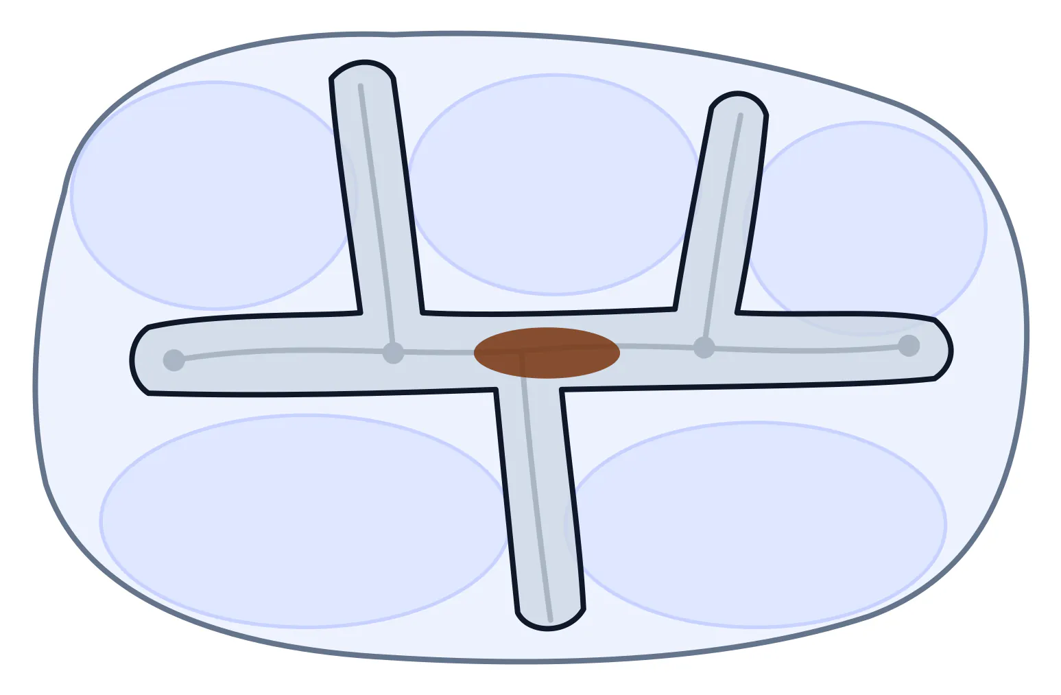
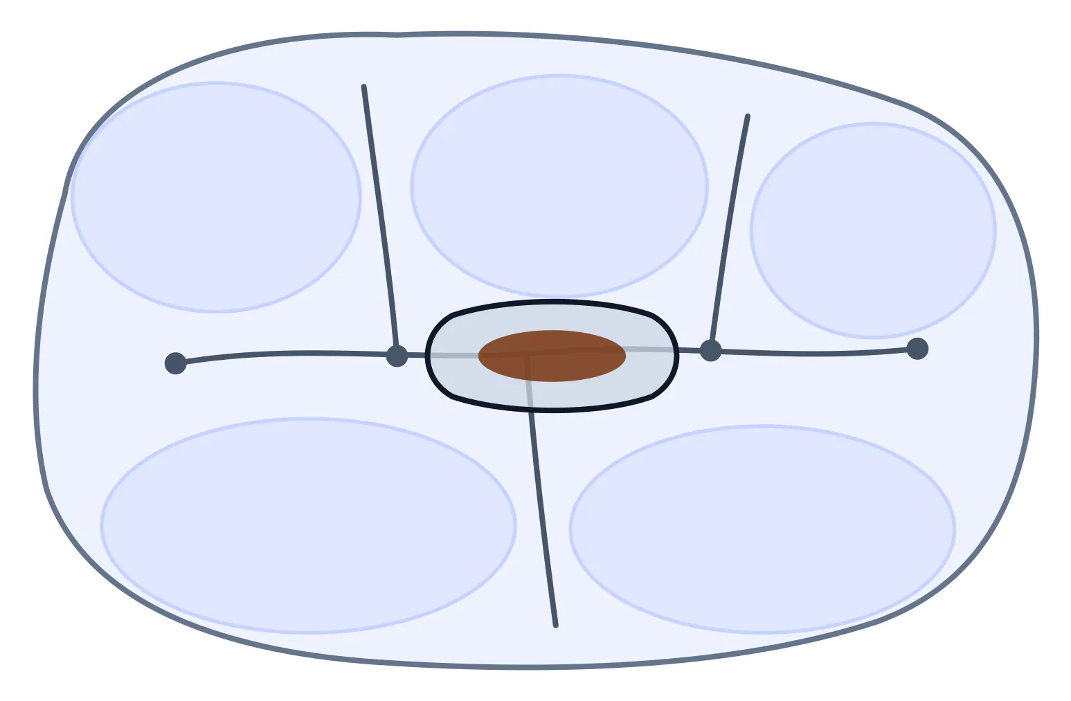
Cavity outline for the same carious lesion in the central fossa of a mandibular molar (diagram, occlusal view): Black's outline with extension for prevention along all fissures (a); minimally invasive outline limited to the lesion, the remaining fissures left intact (b)
Tunnel preparation — Access to proximal caries via the occlusal surface without destroying the marginal ridge. Indicated for initial and dentinal Class II caries in patients with good plaque control.
Slice technique — Proximal cavity preparation using a minimal semicircular slot approach. Preserves significantly more healthy tissue than the classical proximal box.
Air abrasion — Removal of carious tissue by a stream of aluminum oxide particles (27–50 µm) under pressure. Effective for small cavities and fissure caries; often requires no anaesthesia. Limitation: unsuitable for deep cavities.
Chemo-mechanical removal — Gels based on sodium hypochlorite (Carisolv) or papain (Papacárie) chemically soften infected dentin for manual excavation. Minimal invasion, no rotary instruments, often no anaesthesia for shallow lesions.
Summary
Correct cavity preparation is the foundation of a durable restoration. Adhering to the principles — complete caries removal, a flat floor, straight walls, a 90° cavosurface angle and rounded internal angles — ensures a reliable marginal seal and prevents secondary caries. The modern approach calls for maximum preservation of healthy tissue, provided all infected dentin is removed.