Panoramic radiograph — systematic approach and clinical interpretation
ICD-10: Z01.8
OPG: Principle and Clinical Significance
OPG (orthopantomogram) is a panoramic radiograph displaying both jaws, TMJ, maxillary sinuses, inferior alveolar canals and all teeth on a single image.
Clinical value: screening the entire dentition in a single exposure (dose ~0.01–0.03 mSv). Limitation: 2D image with structure superimposition, lower detail than periapical radiograph.
Applications: Screening, treatment planning, implantology — Does not replace periapical radiograph
Dose: ~0.01–0.03 mSv — 2–5× higher than a periapical radiograph
Detail: Lower than periapical radiograph — Caries diagnosis requires periapical/bitewing
Systematic Inspection Zones on OPG
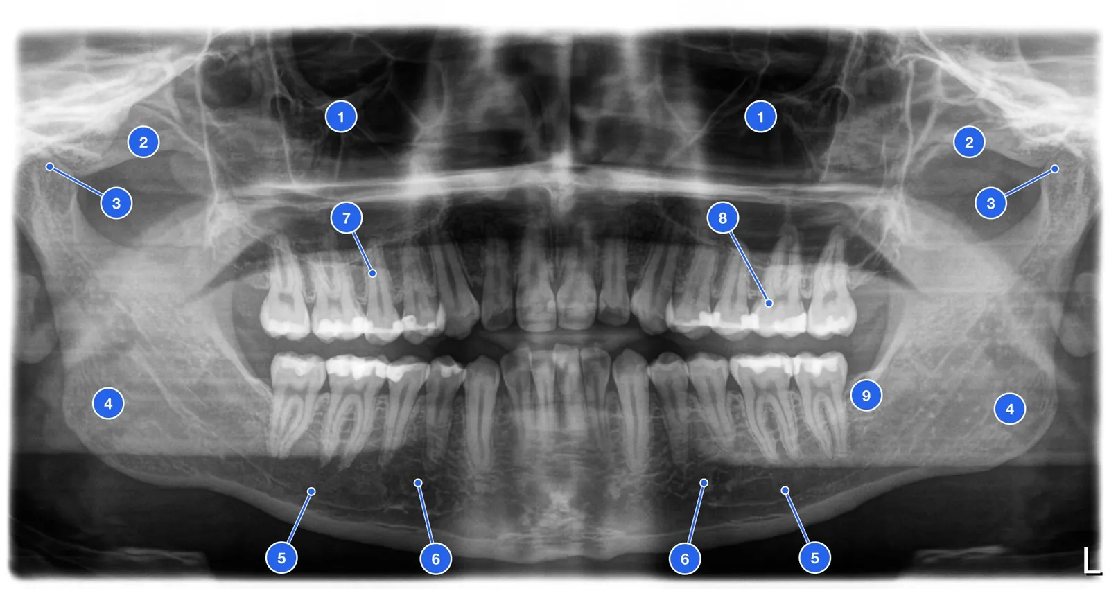
Systematic OPG assessment includes 9 zones. Always examine them in the same order — from the periphery toward the center.
9 OPG inspection zones (sequence):
• 1. Maxillary sinuses (symmetry, pneumatization, opacification)
• 2. Zygomatic bones and arches
• 3. Temporomandibular joints (TMJ): condyles, joint space
• 4. Mandibular rami and angles
• 5. Inferior alveolar canals (n. alveolaris inferior) — right and left
• 6. Mental foramina
• 7. Alveolar ridges (bone resorption, interdental septa)
• 8. Maxillary and mandibular teeth (crowns, roots, interproximal contacts)
• 9. Impacted/supernumerary teeth and pathological lesions
Normal Structures on OPG
Many anatomical structures project onto the OPG and can simulate pathology if the normal anatomy is not known.
| Structure | Appearance | Location |
|---|---|---|
| Inferior alveolar canal | Dark band between two white lines | Mandibular body from retromolar area to mental foramen |
| Maxillary sinus | Dark cavity above upper molar roots | Maxilla — symmetrically on both sides |
| Mental foramen | Round dark spot | Near apex of mandibular 2nd premolar |
| TMJ condyles | Round/oval radiopaque heads | Symmetrically at the tops of the rami |
| Nasal septum | Vertical radiopaque stripe in the midline | Nasal cavity |
Pathological Changes on OPG
Systematic inspection allows detection of pathology in all anatomical zones. Key findings are those that affect treatment planning.
Bone: Diffuse or localized resorption — Periodontitis: horizontal/vertical resorption
Sinuses: Opacification (murkiness) — Sinusitis, polyp, rhinogenic pathology
TMJ: Condylar asymmetry, joint space narrowing — Dysfunction, arthritis, destruction
Impaction: Tooth within bone, unerupted — Wisdom teeth, canines, supernumeraries
Cysts: Round radiolucent lesions — Follicular, radicular, OKC
OPG Limitations and When CBCT Is Needed
OPG is a screening tool. When pathology is found, further evaluation with periapical radiograph or CBCT is often required.
When OPG is insufficient:
• Implant planning in a critical zone → CBCT (distance to canal)
• Suspected vertical root fracture → CBCT
• Assessing root canal anatomy → CBCT
• Caries diagnosis → periapical radiograph / bitewing
• Periapical changes → periapical radiograph
• Wisdom tooth pathology relative to inferior alveolar canal → CBCT
Role of OPG: Overview → screening → plan — Initial diagnostics, not detailed analysis
Detail → periapical radiograph: Caries, periapical tissue — OPG does not replace periapical radiograph