Acute odontogenic inflammation of the periosteum and soft tissues
ICD-10: K10.2 / K04.7
Definition and Epidemiology
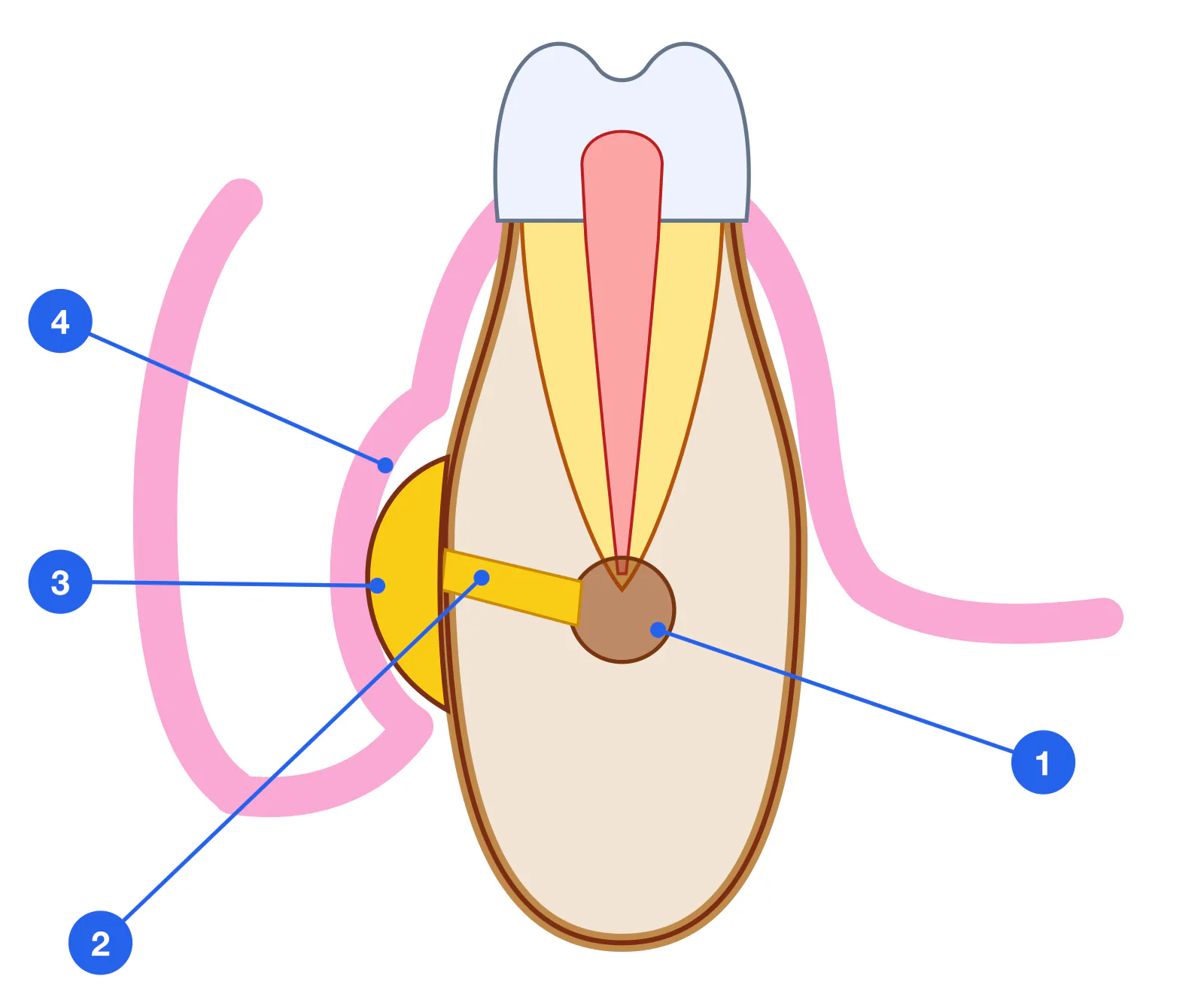
Acute odontogenic periostitis is acute inflammation of the periosteum of the alveolar process, developing as a complication of chronic apical periodontitis or exacerbation of pulpitis. The periosteum is a dense two-layered connective tissue membrane: an outer fibrous layer (vasculo-neural) and an inner cambial (osteogenic) layer bearing osteoblasts. Purulent liquefaction produces a subperiosteal abscess.
An odontogenic abscess is a circumscribed purulent-necrotic focus that forms in the soft tissues (subgingival, submucosal, subcutaneous, subfascial) after infection breaches the cortical plate from a periapical focus.
Odontogenic inflammatory diseases account for 80–85% of all acute maxillofacial pathology. Periostitis is the most frequent form (up to 60% of emergency surgical referrals). Peak incidence: 20–40 years; males are slightly more affected. Most common causative teeth on the mandible: first molars (36, 46); on the maxilla: canines (13, 23) and first molars (16, 26).
Odontogenic proportion: 80–85% — of all acute maxillofacial inflammation
Among surgical emergencies: ~60% — are periostitis cases
EPT in periostitis: > 100 μA — pulp necrosis
Body temperature: 37.5–38.5 °C — in purulent stage
ICD-10: K10.2 / K04.7 — periostitis / periapical abscess
Without treatment: 3–5 days — to progression → phlegmon
Pathogenesis and Spread Anatomy
Pathogenesis of periostitis: chronic apical focus → exacerbation → cortical bone lysis at the apex → infection reaches the subperiosteal space → subperiosteal abscess. Transition from serous to purulent stage takes 24–48 hours with a virulent flora.
The anatomical behaviour of the maxilla versus the mandible is fundamentally different. Maxilla: thin buccal cortex allows easy breakthrough to the vestibule or buccal fat; palatal roots of upper molars direct pus palatally. Mandible: thick cortical plate; the direction of spread is determined by the position of the apex relative to the mylohyoid muscle attachment. Apices of 36–38 lie below m. mylohyoideus → pus tracks into the submandibular space. Apices of 31–35 lie above → submucosal/vestibular abscess.
| Causative tooth | Direction of spread | Anatomical space |
|---|---|---|
| 13, 23 (canines) | Buccally and superiorly | Canine fossa (fossa canina), lower eyelid |
| 16, 26 (palatal roots) | Palatal | Palatal abscess |
| 16, 26 (buccal roots) | Buccally | Buccal space, submasseteric |
| 36, 37 (lower molars) | Below m. mylohyoideus | Submandibular space |
| 31–35 (lower anteriors/premolars) | Above m. mylohyoideus | Vestibular, submental |
Classification
Serous Serous periostitis — Initial stage: hyperaemia, oedema of the periosteum, serous exudate. No fluctuation. Reversible with source elimination and antibiotics within the first 24 h.
Purulent Purulent periostitis — Subperiosteal abscess: purulent liquefaction with stripping of periosteum from bone. Fluctuation, systemic toxicity, trismus. Periostotomy is mandatory.
Diffuse Diffuse periostitis — Pus has spread subperiosteally along the buccal surface across several teeth. Marked collateral oedema. Boundary with phlegmon is indistinct.
Chronic Chronic periostitis — Ossifying or simple. Periosteal thickening with new bone formation (proliferative form). Attenuated clinical picture. Radiograph: "onion skin" periosteal reaction.
Clinical Presentation
Pain — Intense, throbbing, constant. VAS 7–9. Unrelieved by standard NSAIDs. Radiates to temple, ear, orbit.
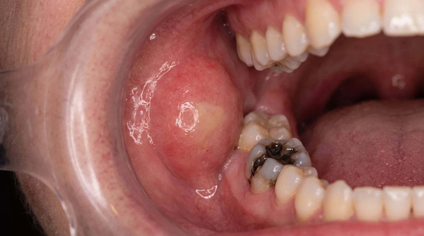
Collateral oedema — Facial asymmetry. Upper jaw: swelling of cheek, lower/upper eyelid, nasal ala. Lower jaw: angle of mandible, submandibular region, neck.
Fluctuation — Bimanual palpation: pressure on one side produces displacement on the other — sign of pus collection. Mandatory periostotomy once fluctuation is present.
Pyrexia — 37.5–38.5 °C in purulent periostitis. Above 38.5 °C — risk of spread (phlegmon, sepsis).
Causative tooth — Broken or previously root-treated. Percussion extremely tender. EPT > 100 μA. Radiograph: widened PDL space, apical bone destruction.
Sulcular reflection — Smoothed out, erythematous, oedematous. In purulent stage — fluctuant swelling along the reflection fold, tender on palpation.
Diagnostics and Differential Diagnosis
| Method | Finding | Note |
|---|---|---|
| EPT (Electric Pulp Test) | > 100 μA — pulp necrosis | Causative tooth non-responsive. Check adjacent teeth for differential diagnosis. |
| Periapical radiograph | Widened PDL space, apical cortical bone destruction, chronic granuloma | Acute changes may not appear radiographically if history is very short. |
| Palpation (fluctuation) | Ballottement sign — indicates formed abscess | Two-finger technique: pressure on one side produces movement on the other. |
| FBC | Leukocytosis 10–15×10⁹/L, left shift, elevated ESR/CRP | Performed in severe cases. Usually not done in outpatient setting. |
| CT / CBCT | Subperiosteal fluid, abscess volume, extent of spread | Indicated when boundaries are unclear or phlegmon is suspected. |
| Feature | Periostitis | Soft tissue abscess | Phlegmon | Exacerbated chronic periodontitis | Acute sinusitis |
|---|---|---|---|---|---|
| Boundaries | Subperiosteal | Localized cavity | Diffuse infiltrate | Apical region only | Maxillary sinus |
| Fluctuation | Present (purulent) | Clear fluctuation | Absent | Absent | Absent |
| Temperature | 37.5–38.5 °C | 37–38 °C | 38.5–40 °C | 37–37.5 °C | 38–39 °C |
| Trismus | Grade I–II | Absent / Grade I | Grade II–III | Absent | Absent |
| Facial asymmetry | Moderate | Localized | Marked | Minimal | Absent |
| Setting | Outpatient | Outpatient | Hospital | Outpatient | ENT + outpatient |
Periostotomy Technique
Periostotomy is an incision through mucosa and periosteum to bone to establish drainage of purulent exudate. It is the cornerstone surgical procedure in purulent periostitis and odontogenic abscess.
Anaesthesia — Infiltration or regional block (mental, Gow-Gates/Vazirani-Akinosi, posterior superior alveolar). Injection into healthy tissues surrounding the inflammatory infiltrate — NOT into the center of the abscess (risk of spread; poor efficacy in acidic environment). For trismus — use regional block technique. (5–10 min)
Incision — Scalpel along the mucosal reflection at the point of maximum fluctuation. Length 1.5–2.0 cm. Incision through mucosa and periosteum in a single motion to bone. Incision direction: parallel to the mucogingival junction (horizontal) to avoid vascular damage. (Scalpel No. 15 or pointed blade) (1–2 min)
Pus evacuation — Blunt dissection in the subperiosteal plane using a periosteal elevator or hemostat. Irrigation with warm 0.9% saline under gentle pressure to flush the cavity. (Periosteal elevator, curved hemostat, irrigation syringe) (2–5 min)
Drainage — Rubber drain (strip of latex glove, 0.5 cm wide) inserted into the wound and secured with a suture to the mucosa for 1–2 days; extend to 3 days for large abscesses. (Rubber drain, silk 3/0) (1 min)
Antiseptic irrigation — Irrigate wound with 0.05% chlorhexidine or 0.01% miramistin. Prescribe rinses every 3 hours for the duration of drainage. (0.05% chlorhexidine, 0.01% miramistin)
Management of causative tooth — Simultaneously with periostotomy or at the same appointment if feasible — extraction or commencement of endodontic treatment (pulpectomy). Leaving the causative tooth untreated is absolutely contraindicated. (Same visit or next day)
Antibiotic Algorithm
Antibiotics in periostitis suppress residual infection after surgical drainage and prevent spread. Antibiotic monotherapy without surgical drainage is ineffective. Drug choice is based on the typical flora of odontogenic abscesses: streptococci, anaerobes (Prevotella, Fusobacterium, Porphyromonas gingivalis).
| Agent | Dose / regimen | Indication | Notes |
|---|---|---|---|
| Amoxicillin/clavulanate | 875/125 mg × 2 /day × 5–7 days | First line | Broad spectrum including anaerobes |
| Amoxicillin/clavulanate | 625 mg × 3 /day | Alternative regimen | Higher daily clavulanate (375 mg vs 250 mg) — more diarrhoea; the 875/125 × 2 regimen is preferred with GI intolerance |
| Clindamycin | 300 mg × 3 /day × 5–7 days | Penicillin allergy | Excellent bone penetration |
| Metronidazole + Amoxicillin | 500 mg × 3 /day + 500 mg × 3 /day | Severe infection, anaerobic cover | Metronidazole contraindicated in the 1st trimester of pregnancy |
| Cefuroxime axetil | 500 mg × 2 /day | Penicillin allergy without history of anaphylaxis (contraindicated after anaphylaxis) | 2nd generation cephalosporin |
| Azithromycin | 500 mg × 1 /day × 3 days | Mild infection, β-lactam intolerance | Growing resistance in oral streptococci |
Complications of Delayed Treatment
Phlegmon of the maxillofacial region — Breakthrough of infection beyond the periosteum into the fascial spaces. Diffuse purulent inflammation without distinct boundaries. Life-threatening.
Osteomyelitis — Purulent inflammation of the bone marrow of the mandible or maxilla. Bone sequestration, sinus tracts, pathological fractures.
Odontogenic maxillary sinusitis — Spread through the floor of the maxillary sinus (roots 15, 16, 17, 25, 26, 27). Unilateral sinusitis with classic dental source.
Mediastinitis — Spread of phlegmon via the visceral cervical space into the mediastinum. Mortality exceeds 40%.
Cavernous sinus thrombosis — Infection tracking from upper teeth along venous pathways. Hectic fever, meningism, exophthalmos — potentially fatal.