Diffuse purulent inflammation of maxillofacial fascial spaces
ICD-10: K12.2
Definition and Fascial Space Anatomy
Maxillofacial phlegmon (cellulitis) is an acute diffuse purulent inflammation of areolar connective tissue — the loose connective tissue filling the spaces between muscles, fasciae and organs — without clear boundaries and with no tendency to self-limitation. The source is odontogenic infection in 80–85% of cases, less often tonsillogenic, traumatic or hematogenous. Microbial flora: polymicrobial associations of streptococci (S. viridans, S. anginosus group), Bacteroides, Prevotella, Fusobacterium and Peptostreptococcus.
Maxillofacial fascial spaces are anatomically divided into superficial (buccal, masseteric, temporal, submental, submandibular) and deep (pterygomandibular, parapharyngeal, sublingual, retropharyngeal, parotid). Between spaces exist real and potential communications — the pathways of purulent spread.
Odontogenic source: 80–85% — of all maxillofacial phlegmons
Mortality (Ludwig): 50–90% — without treatment
IV antibiotics: Co-amoxiclav 1.2 g — IV every 6–8 h — drug of choice (Kazakhstan MoH protocol)
Key complication: Mediastinitis — via paravisceral space
ICD-10: K12.2 — cellulitis and abscess of mouth
Mallampati III–IV: Tracheotomy! — when airway is at risk
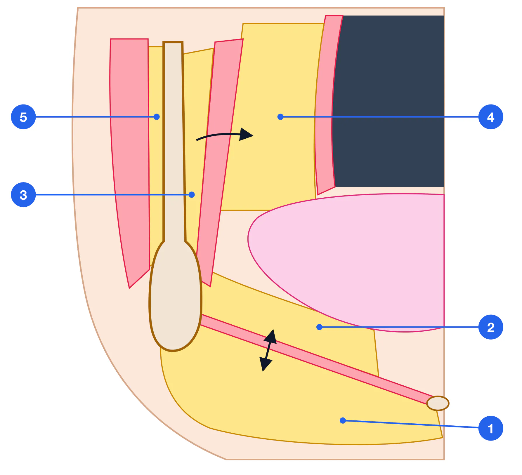
Fascial Spaces: Anatomy and Interconnections
Submandibular Spatium submandibulare — Most frequent location (teeth 36–38, 46–48, whose apices lie below the mylohyoid line). Bounded by m. mylohyoideus (superiorly), platysma and skin (inferiorly). Communicates with sublingual, submental spaces; further spread to parapharyngeal. Incision: skin 2 finger breadths below the inferior border of the mandible.
Sublingual Spatium sublinguae — Above m. mylohyoideus. Source: mandibular incisors, canines and premolars (31–35, 41–45), whose apices lie above the mylohyoid line. Clinical: tongue elevation, hypersalivation, speech impairment. Incision: intraoral, along the lateral floor of mouth parallel to the mandible.
Pterygomandibular Spatium pterygomandibulare — Between the mandibular ramus (lateral) and m. pterygoideus medialis (medial). Source: 38, 48. Trismus is the cardinal sign. Communicates with the parapharyngeal space — gateway to the mediastinum. Incision: extra-oral approach skirting the mandibular angle.
Parapharyngeal Spatium parapharyngeum — Highest risk — direct route to the mediastinum. Clinical: dysphagia, bulging of the lateral pharyngeal wall, oropharyngeal asymmetry, trismus. Mallampati assessment mandatory. Incision: extra-oral along the anterior border of sternocleidomastoid.
Masseteric Spatium massetericum — Between the masseter and the mandibular ramus. Source: 38, 48. Marked trismus; may mimic parotitis. Incision: curved incision at and below the mandibular angle.
Buccal Spatium buccale — Source: upper and lower posterior teeth. Localized cheek swelling, relatively mild course. Incision: intraoral along the sulcus or extraoral when spreading.
| Space | Key sources | Spread pathways | Incision |
|---|---|---|---|
| Submandibular | 36–38, 46–48 | Sublingual, submental, parapharyngeal | Skin 2 finger-breadths below mandible |
| Sublingual | 31–35, 41–45 | Submandibular, pterygomandibular, Ludwig's | Intraoral — floor of mouth |
| Pterygomandibular | 38, 48 | Parapharyngeal → mediastinum | Extraoral, skirting angle of mandible |
| Parapharyngeal | 38, 48, 18, 28 | Retropharyngeal → mediastinum | Along anterior SCM border |
| Masseteric | 38, 48 | Temporal, pterygomandibular | Curved at angle of mandible |
| Buccal | 14–17, 34–37 | Temporal, orbit | Intraoral or extraoral |
Ludwig's Angina
Ludwig's angina is bilateral phlegmonous involvement of the submandibular, sublingual and submental spaces simultaneously. Described by Wilhelm von Ludwig in 1836. It is in fact a distinct clinical syndrome rather than tonsillar angina — surgeons use the term to denote this particular localization.
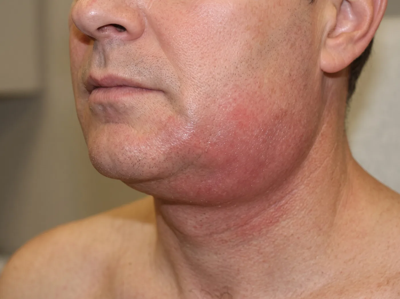
Pathognomonic features: "woody" induration of the infiltrate (putrid-necrotic form with gas-producing anaerobes — clostridia and peptostreptococci), marked tongue elevation with asphyxia risk, characteristic putrid halitosis. Fluctuation is typically absent.
Clinical Presentation and Severity Assessment
Diffuse infiltrate without clear boundaries — Skin over the infiltrate is erythematous, shiny, cannot be pinched into a fold. Fluctuation is uncharacteristic (unlike an abscess) but may appear as liquefaction occurs.
High fever and systemic toxicity — 38.5–40 °C, tachycardia > 100 bpm, RR > 20/min, malaise, confusion. Under the current Sepsis-3 definition (2016), sepsis is not a count of SIRS criteria but life-threatening organ dysfunction caused by infection (an increase in SOFA score ≥ 2 points); for rapid bedside screening use qSOFA: RR ≥ 22/min, altered mentation, systolic BP ≤ 100 mmHg (≥ 2 signs — high risk).
Grade II–III trismus — Mouth opening restricted by masticatory and pterygoid muscle involvement. < 2 cm in grade II, < 1 cm in grade III. Criterion for immediate hospitalization.
Dysphagia and dysarthria — When parapharyngeal and sublingual spaces are involved. Patient cannot swallow saliva; speech sounds like "hot potato voice".
Dyspnea and stridor — Life-threatening! Sign of epiglottis or tongue base oedema. Immediate Mallampati assessment of the airway. Call anaesthetist.
Retrosternal pain (Gerke's sign) — Tilting the head back intensifies retrosternal pain in mediastinitis — that is the Gerke sign; the forced position is the opposite, with the chin held towards the chest. Dyspnea, widened mediastinum on chest radiograph. CT chest — immediately.
| Feature | Abscess | Phlegmon | Ludwig's Angina |
|---|---|---|---|
| Boundaries | Distinct | Indistinct | Indistinct, bilateral |
| Fluctuation | Present | Usually absent | Absent (woody induration) |
| Temperature | 37–38 °C | 38.5–40 °C | 39–40 °C |
| Trismus | None / Grade I | Grade II–III | Grade III (often < 1 cm) |
| Airway risk | None | Rare | YES — primary risk |
| Setting | Outpatient | Hospital | ICU + tracheotomy |
Diagnostics
| Method | Finding | Note |
|---|---|---|
| FBC | Leukocytosis > 15×10⁹/L, left shift, neutrophilia, ESR > 40 mm/h | In sepsis: WBC < 4 or > 12, toxic granulation may be present. |
| CRP, procalcitonin | CRP > 100 mg/L; PCT > 2 ng/mL → sepsis | PCT < 0.5 — bacteremia unlikely. |
| Coagulation screen | INR, APTT — assess for DIC in severe sepsis | Thrombocytopenia + prolonged PT → DIC. |
| Blood cultures | 2 aerobic/anaerobic pairs before antibiotics | Positive in 20–40% of phlegmon cases. |
| CT neck with contrast | Ring enhancement = abscess; diffuse = cellulitis; gas = anaerobes | Investigation of choice. Defines extent, guides surgery. |
| CT chest | Widened mediastinum, pleural effusion → mediastinitis | Mandatory when mediastinitis suspected (retrosternal pain, Gerke's sign). |
| Mallampati assessment | Class I–II: intubation feasible; III–IV: consider tracheotomy | Performed for any phlegmon with trismus or floor-of-mouth oedema. |
Surgical Management
Emergency admission — To the maxillofacial surgery unit. Airway at risk — direct to ICU. Anaesthetist to be called for any trismus above grade I. (Immediately)
Stabilization and investigation — FBC, metabolic panel, coagulation screen, blood cultures × 2 (before antibiotics). CT neck and chest. Mallampati assessment. SpO₂ monitoring. IV access × 2. (30–60 min)
Airway management — Awake fiberoptic intubation — when Mallampati III–IV or trismus < 2 cm. Tracheotomy under local anaesthesia — when floor of mouth severely oedematous or intubation impossible. NEVER mask induction when airway is at risk. (Tracheotomy set MUST be in theatre and ready)
Wide incision and drainage — Principle: open ALL involved spaces. Incisions: external (submandibular, "collar incision" for Ludwig's) + intraoral. For mediastinitis — VATS or thoracotomy (thoracic surgeon). (Scalpel, clamps, diathermy)
Drainage — Active drainage through multiple incisions. Drains: rubber tubing, penrose drains, NPWT (negative pressure wound therapy) for extensive necrotic phlegmon. Daily dressing changes and irrigation. (Drain tubing, 20 mL syringes, 0.9% NaCl + miramistin)
Extraction of causative tooth — At earliest opportunity under antibiotic cover. In the acute phase of Ludwig's angina — deferred until patient is stabilized. (Within 24–48 h if feasible)
Repeat wound review — Daily assessment: wound spread, drain function. Repeat CT on clinical deterioration or at 48–72 h post-operatively. (Every 24 h)
In-Hospital Antibiotic Regimens
| Regimen | Agents | Dose / schedule | Indication |
|---|---|---|---|
| Drug of choice (LoE A) | Co-amoxiclav | 1.2 g IV every 6–8 h, 7–10 days | Cellulitis and abscess of the mouth |
| Alternative (LoE B) | Lincomycin | 0.6–1.2 g IV infusion every 12 h, 7–10 days | When the drug of choice is unsuitable |
| Pseudomonas infection | Ceftazidime or ciprofloxacin | Ceftazidime 3–6 g/day in 2–3 doses; ciprofloxacin 0.4–0.6 g IV infusion every 12 h | P. aeruginosa on culture |
| Sepsis (outside the protocol) | Piperacillin/tazobactam | 4.5 g IV × 4/day | Sepsis (Sepsis-3), multiple organ failure (MOF), mediastinitis — per international practice, together with the intensivist |