Pathophysiology, classification, treatment protocol and prevention
ICD-10: K10.3
Definition and Pathophysiology
Alveolitis (alveolar osteitis, dry socket) is inflammation of the walls of an extraction socket resulting from failure of blood clot formation or lysis of the clot by fibrinolytic enzymes. It is characterized by exposure (necrosis) of the socket bone, intense pain and halitosis.
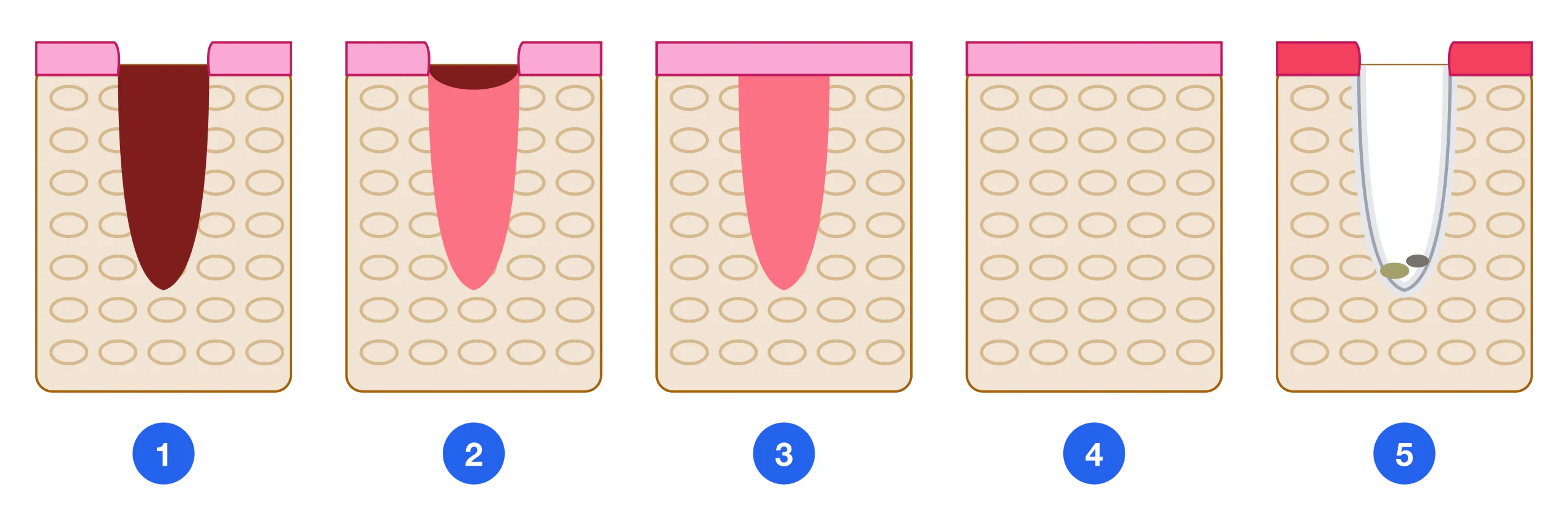
Pathophysiological mechanism: normal socket healing — fibrin clot formation (1–2 h) → organization by granulation tissue (3–7 days) → epithelialisation (7–14 days) → mineralization (3–6 months). In alveolitis: premature clot destruction due to activation of tissue plasminogen activator (t-PA) → fibrinolysis → bone exposure → secondary bacterial contamination → inflammation. Role of nicotine: vasoconstriction reduces local blood flow and retards clot formation; suction creates negative intra-socket pressure that mechanically dislodges the clot.
Epidemiology: overall incidence 2–5%, rising to 20–30% after mandibular third molar extractions (38, 48). Onset: day 3–5 post-extraction. Pain VAS 7–9, unresponsive to standard NSAIDs. Without treatment, an unhealing socket carries a risk of progression to osteomyelitis.
Overall incidence: 2–5% — of all extractions
After 38/48: 20–30% — lower third molars
Symptom onset: Day 3–5 — post-extraction
Pain VAS: 7–9 — unresponsive to NSAIDs
Estrogen (OCP): ↑ risk — enhances fibrinolysis
ICD-10: K10.3 — alveolitis of jaw
Risk Factors
Risk factors for alveolitis
Modifiable behavioural: Smoking (primary factor), Rinsing within 24 h, Drinking through a straw, Hot food/drinks
Systemic factors: Oral contraceptives (estrogen), Diabetes mellitus, Immunosuppression, Coagulation disorders
Local / technical: Traumatic extraction, Active pericoronitis before extraction, Deep impaction, Chronic periodontitis
Pharmacological: Anticoagulants (heparin, warfarin), Corticosteroids, Bisphosphonates (MRONJ risk)
Classification
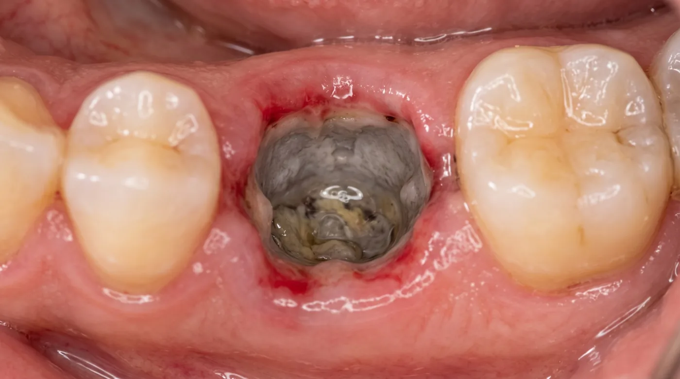
Dry socket Fibrinolytic alveolitis — Complete clot loss. Socket empty — no clot, no granulation tissue. Exposed bone is grey-white, tender on probing. Halitosis. Pain intense (VAS 8–9), radiating to ear and temple. Day 3–5.
Infected Bacterial alveolitis — Clot present but necrotic — grey, malodorous. Moderate pain. Erythema of socket margins, regional lymphadenitis.
Partial Partial (localized) — Some socket walls covered with granulation tissue, others exposed. Intermediate form.
Generalised Generalised form — Multiple wall involvement, risk of osteomyelitis. Fever, marked lymphadenitis. Systemic antibiotics required.
Differential Diagnosis
| Feature | Normal pain (day 1–2) | Alveolitis (day 3–5) | Osteomyelitis (week 1+) | Sinusitis |
|---|---|---|---|---|
| Pain onset | Immediately after extraction | Day 3–5 (worsening) | Week 1+ | Several days |
| Pain character | Moderate, subsiding | Intense, not subsiding | Constant + dull | Pressure in sinus area |
| Socket | Dark red clot | Empty / grey breakdown | Sequestrum (later) | No change |
| Temperature | Normal | < 37.5 °C (if not infected) | > 38 °C | 38–39 °C |
| NSAID effect | Good | Minimal | Weak | Moderate |
| Radiograph | Normal | Normal / indistinct walls | Sequestra, destruction | Sinus opacity |
Treatment Protocol
Anaesthesia — Regional block or infiltration. Do not inject into the zone of inflammation (reduced efficacy due to acidosis). For lower teeth: inferior alveolar or mental nerve block. (5 min)
Socket irrigation — Irrigate with warm (37 °C) 0.9% saline via a blunt-tipped needle or Moult cannula (reverse-flow technique). Moderate pressure — avoid dislodging any remaining granulation tissue. Aspirate necrotic debris gently. Volume: 10–20 mL per session. (0.9% saline, 0.05% chlorhexidine, 0.01% miramistin, 20 mL syringe, 18G blunt needle) (3–5 min)
Gentle curettage (if indicated) — Only when frank necrosis is present at the socket floor. Careful, without traumatising bone. Do NOT perform "socket re-freshening" (curettage to bleeding) — this is an outdated method that increases inflammation. (Small curette)
Socket dressing — Alvogyl: combination of butamben (anaesthetic) + eugenol (antibacterial) + iodoform (antiseptic). Pack loosely into the socket, do not compress deeply. Alternatives: ZnO-eugenol paste, BIPP (bismuth-iodoform-paraffin paste). Analgesic effect: within 10–15 minutes. (Alvogyl, ZnO-eugenol paste, college tweezers, sterile gauze strip) (2–3 min)
Systemic analgesia — Ibuprofen 400–600 mg × 3/day (first-line NSAID). For severe pain: ketorolac 10–30 mg or nimesulide 100 mg. Paracetamol 500 mg × 4/day — adjunct. (3–7 days)
Antibiotics — For infected alveolitis or signs of spread (lymphadenitis, temperature > 37.5 °C): amoxicillin 500 mg × 3/day × 5–7 days, or co-amoxiclav 625 mg × 3/day × 5–7 days. NOT indicated for simple dry socket without infection. (5–7 days)
Repeat dressings — Daily or every other day until granulation tissue appears (typically 5–10 days). Each visit: irrigation + dressing change. Once granulations appear — no further dressing needed. (5–10 days)
Prevention
Pre-operative measures — When active pericoronitis is present — resolve inflammation first (7–10 days antibiotics) before extraction. OCP patients: schedule in the first week of the cycle. Full oral hygiene assessment before elective extraction. (Before the operation)
Atraumatic extraction technique — Minimise trauma to mucosa and bone. Avoid excessive pressure on the socket, limit vasoconstrictor volume. For difficult extractions — prefer bone removal with a bur over forceful elevation. (During the operation)
Local hemostatic measures — Where possible — place a resorbable hemostatic sponge (Gelfoam, Surgicel) into the socket. Sutures over the wound reduce alveolitis risk after complex extractions. (Immediately post-extraction)
Written patient instructions — Do NOT rinse for 24 hours (preserve the clot!). Do NOT smoke for 48–72 h (ideally 1 week). No drinking through straws. Cold pack to cheek for first 24 h. Soft diet. Do not probe socket with tongue. Contact the clinic if pain worsens after day 2. (Written instructions given!)