Indications, contraindications, instruments, steps and complication management
ICD-10: —
Indications and Contraindications
Absolute indications Absolute indications for extraction — Pulp gangrene when endodontic treatment is not feasible; periodontitis grade III–IV with mobility ≥ III; malignancy in the tooth region; tooth as the source of odontogenic infection (periostitis, phlegmon, osteomyelitis) when conservative treatment is impossible.
Relative indications Relative indications — Orthodontic (space creation); pre-prosthetic (tooth obstructing prosthetic plan); prophylactic before radiotherapy; impaction with complications (pericoronitis, cyst).
Absolute contraindications Absolute contraindications — Acute MI or stroke < 6 months; hemorrhagic diatheses (INR > 3.5); malignant tumour at the surgical site; irradiated field (risk of osteoradionecrosis).
Relative contraindications Relative (require preparation) — Anticoagulants — target INR ≤ 2.5–3.0 (do NOT stop warfarin); pregnancy — second trimester preferred; diabetes — HbA1c ≤ 9% before elective extraction; bisphosphonates — MRONJ risk requires risk assessment.
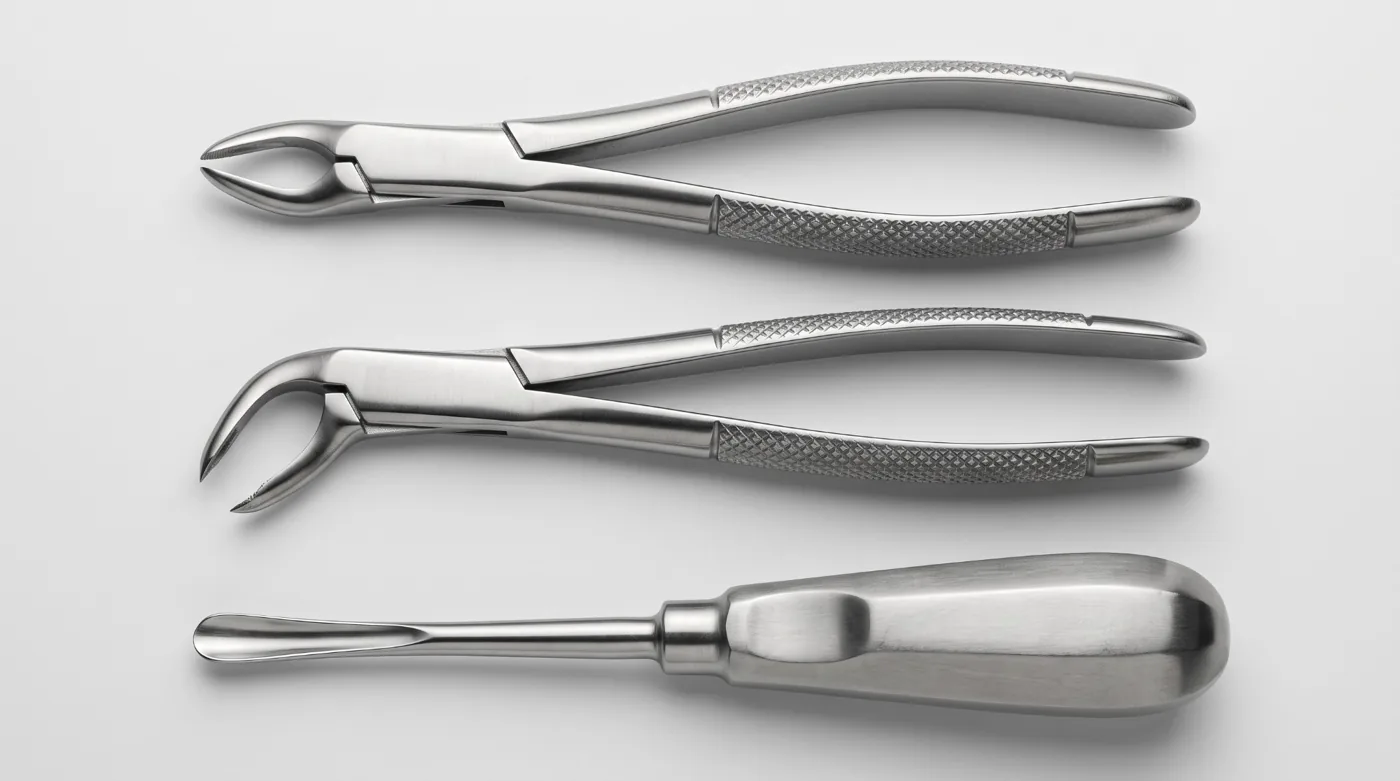
Instruments
| Instrument | Application | Notes |
|---|---|---|
| Upper central incisor forceps | 11, 21 | Straight; conical beaks for single, cylindrical roots |
| Upper canine forceps | 13, 23 | Elongated beaks, stronger grip |
| Upper premolar forceps | 14, 15, 24, 25 | S-curved shanks, narrow beaks |
| Bayonet forceps (upper molars) | 16, 17, 26, 27 | S-shaped with buccal beak — separate left and right |
| Lower universal forceps | 31–37 | Angled at the beak–shank junction; beaks parallel to tooth axis |
| Lower molar forceps (horizontal) | 36, 37, 46, 47 | Horizontal beaks with spikes for the furcation |
| Straight elevator (Coupland) | All teeth | Inserted into PDL space; severs PDL fibers — luxation |
| Angled elevator (Potts) | 38, 48 | Left/right pair for lower wisdom teeth |
| Lecluse elevator | Lower third molars (38, 48) | Inserted between the second and third molar, displaces the third molar distally and upwards |
Extraction Technique
Anaesthesia — Upper teeth: infiltration + palatal block; for molars — posterior superior alveolar block (16–18, 26–28). Lower teeth: inferior alveolar block + mental/buccal long nerve or Gow-Gates technique. 4% articaine with 1:100,000 adrenaline — standard. Onset: 3–5 min infiltration, 5–10 min block. (5–10 min exposure)
Syndesmotomy — Sever the circular (circumdental) gingival fibres at the tooth neck — not the periodontal ligament — with a scalpel or syndesmotome — around the entire circumference of the neck. Essential to prevent tearing the gingiva as the forceps are advanced. (Syndesmotome or scalpel) (1–2 min)
Forceps placement — Forceps axis parallel to the tooth axis. Beaks advanced as far subgingivally as possible (close to bone). Buccal beak first. Secure grip — moderate pressure, no fracture. (30 s)
Luxation — Buccal-palatal (labial-lingual) movements — increasing amplitude, expanding the socket. For single-rooted teeth with a round root cross-section (upper central incisors, canines) — add rotation. Lower incisors (root flattened mesiodistally) and molars: luxation only, no rotation (risk of root fracture). (1–3 min)
Traction — After adequate luxation — smooth delivery along the tooth axis. Upper teeth: downwards and outwards. Lower teeth: upwards and outwards. Avoid sudden jerking movements. (10–30 s)
Socket inspection — Inspect socket: root fragments, granuloma, cyst (requiring curettage), sharp bony edges (to be smoothed). Curette granulation tissue only when an apical granuloma is present — prevents cyst formation. (Curette, hemostatic clamp) (2–3 min)
Hemostasis — Patient bites on gauze pack for 15–20 minutes. Persistent bleeding: suture the socket, add hemostatic sponge (Surgicel, Gelfoam), topical thrombin. Systemic cause (anticoagulants) — systemic correction. (Gauze, Surgicel, Gelfoam) (15–20 min)
Upper Molar Extraction: Risk of OAC
Oroantral communication (OAC) is a pathological opening between the oral cavity and the maxillary sinus, occurring during extraction of upper posterior teeth (most often 16, 17, 26, 27). Incidence: 0.5–3.5%. Risk is higher with: long divergent molar roots, excessive sinus pneumatization, periapical periodontitis of the relevant tooth.
Diagnosis: Valsalva test (patient occludes the nose and gently exhales — air exits through the socket), fluid draining from the nose during rinsing, aspiration of air in the socket. Management: ≤ 2 mm — conservative (no smoking, decongestant nasal drops); 2–5 mm — figure-of-8 suture across the socket; > 5 mm — flap repair (buccal mucoperiosteal advancement flap — Wassmund technique, or buccal fat pad flap). OAF (oroantral fistula) = OAC persisting > 3 weeks → surgical closure mandatory.
Lower Third Molar Extraction (38/48): Risks
The lower third molar (38/48) is the most technically demanding extraction. The principal risk is injury to the inferior alveolar nerve (IAN), which provides sensation to half of the lower lip, mandibular teeth and chin. Incidence of IAN paresthesia: 0.5–5%; the majority resolve within 6–18 months. The lingual nerve (n. lingualis) is at risk if the lingual flap is raised carelessly (paresthesia of the anterior 2/3 of the tongue).
Radiographic assessment: OPG — signs of IAN proximity: root darkening in the canal projection, canal deflection at the point of contact, interruption of the canal cortical line. When uncertain — CBCT for three-dimensional assessment. Technique choice: for shallow impaction — elevator/forceps extraction; for deep impaction — tooth sectioning with a bur (crown + roots separately), ostectomy for access.
Incision and flap reflection — Angular incision (posterior-inferior from 37, along the anterior ramus border + horizontal along the buccal sulcus). Mucoperiosteal flap reflected with a periosteal elevator. Lingual nerve protection — lingual retractor must NOT exert pressure on the lingual bone. (Scalpel, periosteal elevator, Muller retractor)
Ostectomy — Remove bone overlying the crown and distally using a round or fissure bur with copious saline irrigation. Avoid overheating bone (risk of osteonecrosis). Expose down to the cemento-enamel junction. (Surgical handpiece, fissure bur, saline irrigation)
Tooth sectioning — For mesioangular and horizontal impactions — section through the crown at the furcation level. Remove crown and roots sequentially with elevator. If two independent roots — separate them from each other. (Fissure bur, angled Potts elevator)
Inspection and closure — Inspect and curette the socket, ensure hemostasis. Close the flap with 2–3 interrupted sutures. Horizontal mattress suture for a large defect. Vicryl or silk 3/0–4/0. (Vicryl 3/0, needle driver, scissors) (5–10 min)
Complications
| Timing | Complication | Cause | Management |
|---|---|---|---|
| Intra-operative | Crown/root fracture | Ankylosis, technique error | Elevator; bur for access if needed |
| Intra-operative | Hemorrhage | Arterial, coagulopathy | Ligation, suturing, hemostatic agents |
| Intra-operative | Maxillary tuberosity fracture | Ankylosed 17, 27 attached to tuber | If mobile bone: retain and suture; if detached: remove |
| Intra-operative | Displacement of adjacent tooth | Technique error | Reposition + splinting |
| Intra-operative | OAC (sinus perforation) | Long roots 16, 17, 26, 27 | Immediate flap repair if > 5 mm |
| Early post-op (1–7 days) | Alveolitis | Clot lysis | Irrigation, alvogyl, dressings |
| Early post-op | Hematoma | Soft tissue bleeding | Rest, ice; if enlarging — explore |
| Late (> 1 week) | IAN paresthesia | IAN trauma during 38/48 | Observe up to 6 months, vitamin B12 |
| Late | OAF (oroantral fistula) | Untreated OAC | Surgical flap closure |