Andreasen–WHO classification, management of fractures and luxations, replantation protocol
ICD-10: S02.5 / S03.2
Introduction: Scope and Core Principles
Dental trauma is acute mechanical injury to the hard tissues of the tooth, pulp and/or supporting structures (periodontal ligament, alveolar bone, gingiva). It ranks second in frequency of paediatric dental emergency presentations after caries. The clinician's primary management within the first 30–60 minutes fundamentally determines the prognosis of the tooth.
Prevalence: up to 25% of children aged 6–12 sustain trauma to permanent teeth; peak incidence at 8–10 years. In adults: road traffic accidents, sport, assault. Most vulnerable teeth: maxillary central incisors (up to 80% of all dental injuries), particularly with overjet > 4 mm.
Standard of care: International Association of Dental Traumatology (IADT) Guidelines 2020. WHO classification revised by Andreasen (1994) is the reference framework. The clinical Ellis classification is of historical use and does not cover luxations.
Peak incidence: 8–10 years — mixed dentition + physical activity
Most vulnerable: 80% — maxillary central incisors
Critical window: 30 min — avulsion replantation
Gold standard: IADT 2020 — international guidelines
Storage medium: Milk — if no HBSS — viable up to 6 h
EPT errors: Up to 65% — false negative immediately post-trauma
Classification: Hard Tissue Injuries
The Andreasen classification (WHO ICD-DA) divides injuries into two major groups: injuries to the hard dental tissues and injuries to the supporting tissues (luxations). Every injury type must be coded precisely at the initial examination.
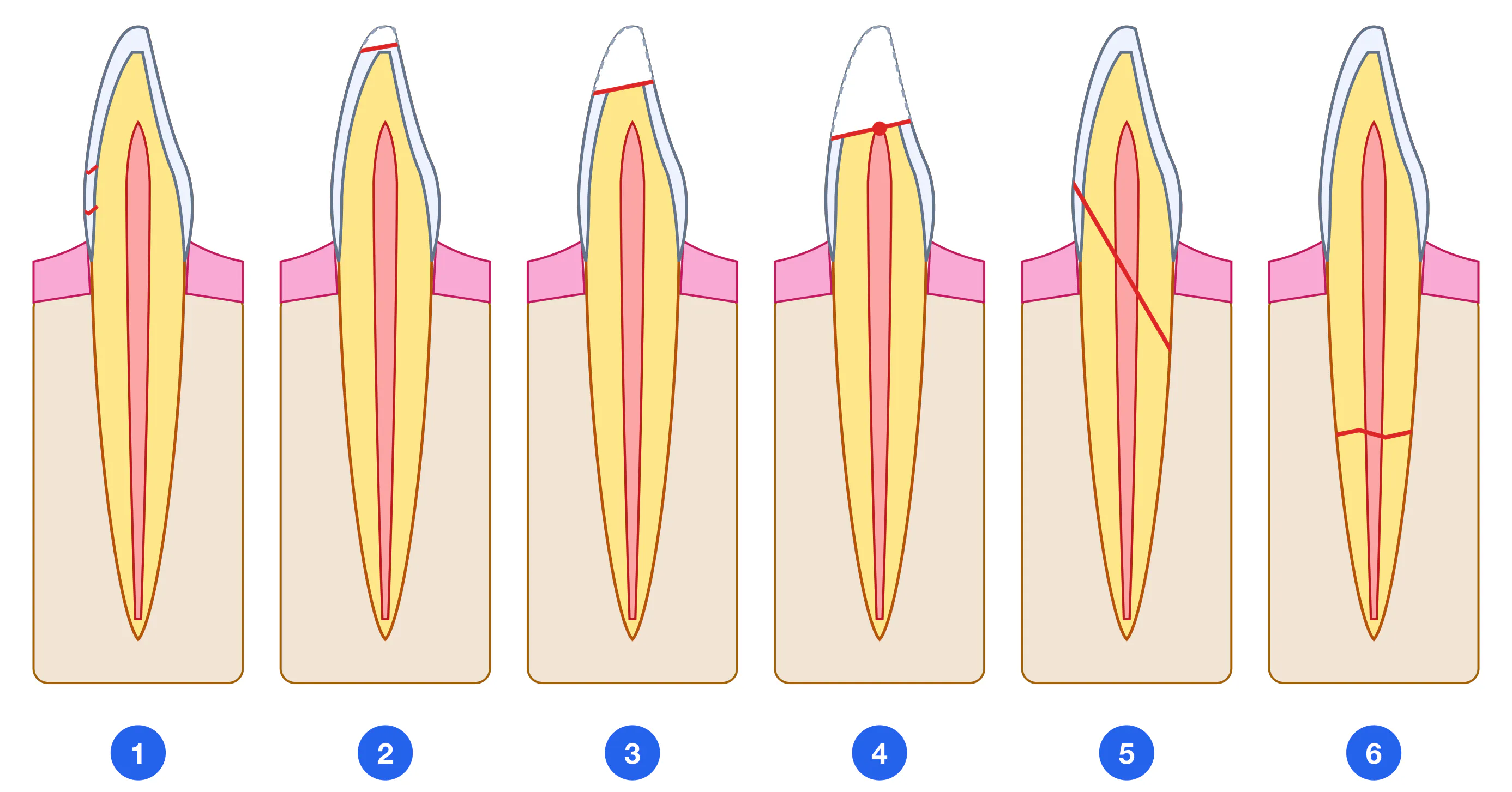
S02.5 Enamel infraction (crack) — Incomplete fracture without loss of tooth substance. Visible on transillumination. Minimal symptoms. Management: smooth sharp edges, fluoride, recheck at 4 weeks.
S02.5 Uncomplicated enamel fracture (Ellis I) — Loss of enamel only. No or minimal pain. EPT normal (2–12 μA). Management: smooth edges + composite restoration or reattachment.
S02.5 Uncomplicated crown fracture (Ellis II) — Enamel + dentin, no pulp exposure. Temperature sensitivity. Management: immediate dentin coverage with calcium-based liner + restoration. EPT at 4, 8, 12 weeks.
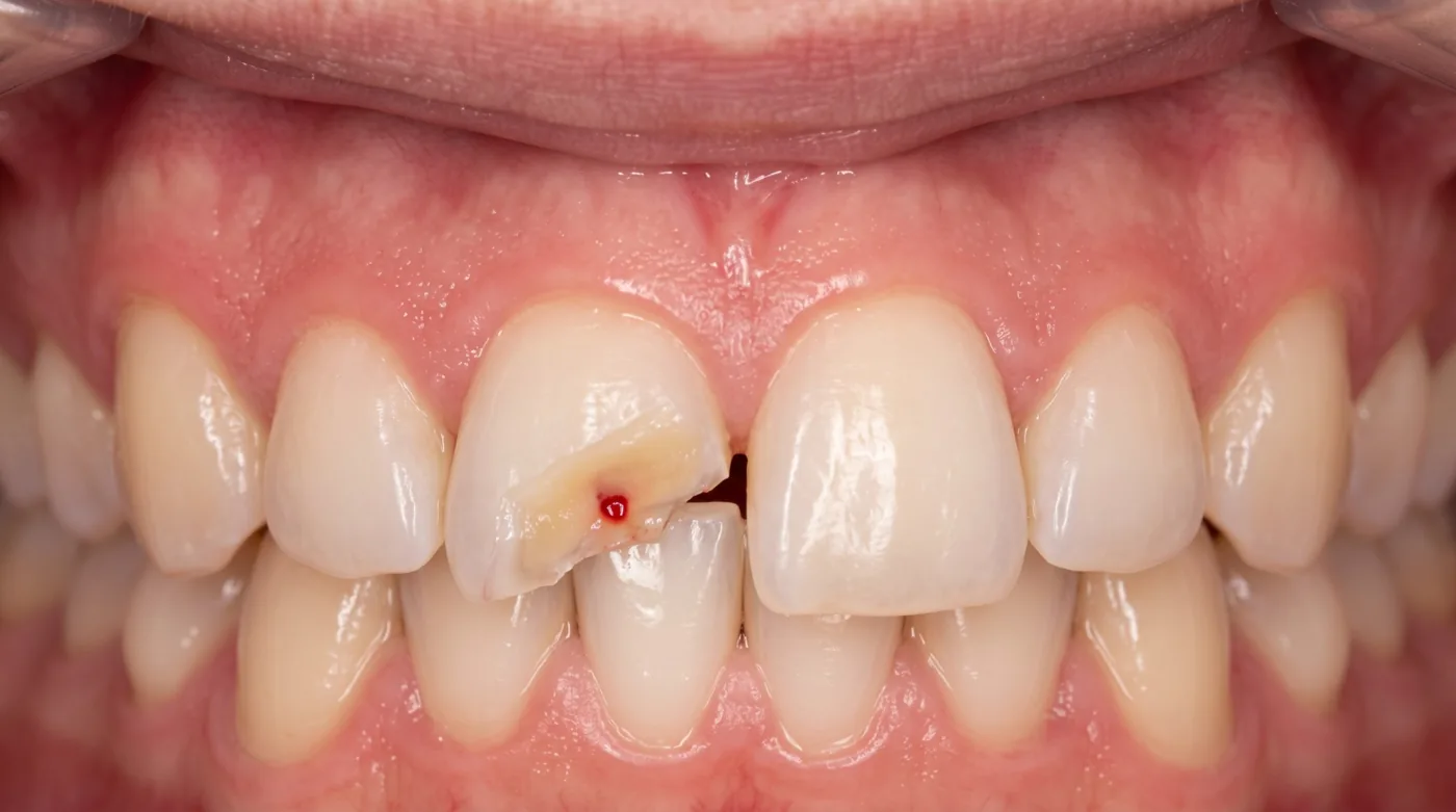
S02.5 Complicated crown fracture (Ellis III, pulp exposure) — Enamel + dentin + pulp exposure (pink spot). Acute pain. Management: immature apex → direct pulp cap (MTA/Biodentine) or pulpotomy; mature apex → pulpectomy + root canal treatment.
S02.5 Crown-root fracture — Fracture line through enamel, dentin and root cementum. Mobile fragment retained by periodontal fibers. CBCT mandatory. Management: remove mobile fragment → assess subgingival level → surgical/orthodontic extrusion or extraction.
S02.5 Root fracture — Fracture line within the root (apical, middle or cervical third). Mobility of coronal fragment. Dull percussion note. Diagnosis: periapical radiographs in 3 projections, CBCT if needed. Management: repositioning + flexible splint 4 weeks (middle/apical ⅓, possible self-healing) or up to 4 months (cervical ⅓) — per IADT 2020.
Classification: Luxation Injuries
Luxation injuries involve damage to the supporting periodontal structures with or without hard tissue injury. The splint must always be flexible (semi-rigid) — rigid splinting impairs PDL healing and promotes ankylosis.
S03.2 Concussion — No displacement, no increased mobility. Tender to percussion. EPT may be false-negative. Management: no splint, soft diet × 2 weeks, monitor EPT at 4, 8, 12 weeks, 6 months, 1 year.
S03.2 Subluxation — Increased mobility, no displacement. Sulcular bleeding. Management: flexible splint × 2 weeks. Prognosis: vitality preserved in ~75% with closed apex.
S03.2 Extrusive luxation (partial avulsion) — Tooth partially displaced out of the socket in a coronal direction along its long axis (appears elongated), grade II–III mobility. EPT negative. Management: repositioning under LA → flexible splint × 2 weeks → endodontics at 4 weeks if non-vital.
S03.2 Lateral luxation — Displacement in vestibular, lingual or distal direction with alveolar bone fracture. Metallic percussion note. EPT negative. Management: repositioning (± surgical decompression) + flexible splint × 4 weeks → endodontics if necrosis.
S03.2 Intrusive luxation (intrusion) — Tooth driven apically into the socket — appears shortened. Metallic percussion note (ankylosis-like). EPT negative. Worst prognosis. Management: immature apex → await spontaneous re-eruption; mature apex, intrusion < 3 mm → wait; > 3 mm → orthodontic extrusion or surgical repositioning + flexible splint 4–8 weeks. Endodontics mandatory for closed apex within 2–3 weeks.
S03.2 Avulsion (complete displacement) — Tooth completely displaced from the socket. Extra-alveolar time and storage medium determine prognosis. Endodontic treatment mandatory for closed-apex teeth at 7–10 days post-replantation.
Diagnostic Algorithm in Dental Trauma
Diagnosis of dental trauma is a structured process that allows precise classification of the injury and selection of management. It is essential not to miss associated injuries (traumatic brain injury, jaw fracture, root fracture).
Trauma history: time, mechanism, storage medium of the tooth (in avulsion) — Loss of consciousness, amnesia, vomiting → rule out TBI. Malocclusion → rule out jaw fracture.
Examination: type and depth of fracture, presence of a pink spot (pulp exposure) — Pink spot = pulp exposure = complicated fracture. Pinkish discoloration without a spot = pulp haematoma (tooth still vital).
Assessment of displacement and mobility: direction, degree (I–III), metallic vs normal percussion note — Metallic percussion note = ankylosis (intrusion, late replacement resorption).
EPT (electric pulp testing): immediately after injury and at 4, 8, 12 weeks, 6 months, 1 year — Normal: 2–12 μA. Immediately after trauma 30–65% of teeth give a false-negative response (pulpal shock, oedema). Only serial EPT + radiographs provide a reliable assessment.
Radiography: periapical radiographs in 2–3 projections, CBCT if root fracture is suspected — CBCT allows 3D assessment of root fracture, degree of intrusion and the state of the alveolar process. Conventional radiographs may miss a horizontal root fracture.
Transillumination: to detect enamel cracks (infractions) — Cracks appear as dark lines under lateral illumination. Untreated infractions carry a risk of caries and chipping.
Replantation Protocol for Avulsion
Avulsion is the only situation in dentistry where the patient or bystanders must begin treatment before reaching the dentist. The viability of periodontal ligament (PDL) cells determines the success of replantation. In dry storage PDL cells die within 15–20 minutes; in milk they survive up to 6 hours.
| Storage Medium | PDL Cell Survival | Availability | Comment |
|---|---|---|---|
| HBSS (Hanks Balanced Salt Solution) | 24+ hours | Pharmacy / school kits | Gold standard — isotonic, pH-buffered |
| Cold milk | up to 6 hours | High | Best practical option; skimmed slightly better |
| Normal saline 0.9% NaCl | 2–6 hours | High | Acceptable when milk unavailable |
| Saliva (buccal vestibule) | 30–120 min | Always available | Infection risk; buccal sulcus preferred over hand |
| Tap water | < 20 min | High | PROHIBITED — hypotonic, lyses PDL cells |
| Dry storage | < 15–20 min | — | Critically poor; > 60 min dry → ankylosis inevitable |
Step 1 — First aid at the scene (patient / bystander) — Pick up tooth by crown (NOT root). Do not scrape or wipe. Rinse gently with saline or milk for 10 seconds. Place in milk or buccal sulcus. Go to dentist immediately. (Milk / saline / HBSS) (< 5 minutes)
Step 2 — Assessment on arrival — Extra-alveolar time and storage medium? Open or closed apex? TBI, fractures? Condition of socket (clot / granulation / contamination)? (5–10 minutes)
Step 3 — Tooth preparation — Tooth < 60 min in liquid: gently rinse root with saline. Tooth > 60 min dry: PDL cells are non-viable and the expected outcome is replacement resorption (ankylosis); soaking the root in sodium fluoride is not part of the IADT 2020 guidelines. NEVER scrub the root! (Saline) (5–10 minutes)
Step 4 — Replantation — Local anaesthesia (infiltration / block). Irrigate socket with saline — do not curette. Place tooth into socket with gentle finger pressure in correct axial orientation. Check occlusion. (Articaine 4%) (5–10 minutes)
Step 5 — Splinting — Flexible splint: orthodontic wire + composite or proprietary splint strip. Splint spans the replanted tooth + 1–2 sound teeth each side. Duration: 2 weeks (IADT 2020). Rigid splint — CONTRAINDICATED (prevents PDL healing, promotes ankylosis). (Composite + orthodontic wire) (15–20 minutes)
Step 6 — Medical treatment — Antibiotics: amoxicillin 500 mg × 3/day × 7 days (children — weight-based dose). Penicillin intolerance: azithromycin 500 mg/day × 3 days. Tetanus prophylaxis (tetanus toxoid 0.5 mL) for contaminated wounds if the last vaccination was more than 5 years ago. NSAIDs: ibuprofen 400 mg. (Amoxicillin, ibuprofen, TT toxoid) (Prescriptions at visit)
Step 7 — Endodontic treatment (closed apex teeth) — At 7–10 days post-replantation (before splint removal). Debridement, temporary Ca(OH)₂ dressing for minimum 1 month (suppresses inflammatory resorption). Permanent obturation at 6–12 months. Open-apex teeth: monitor for revascularisation. (Ca(OH)₂, files, sealer) (45–60 minutes)
Step 8 — Follow-up — Review schedule: 2 weeks (remove splint), 1 month, 3 months, 6 months, 1 year, then annually. Assess: mobility, percussion note, EPT, radiograph (resorption, periapical status). (Scheduled)
Splinting Duration by Injury Type (IADT 2020)
| Injury Type | Splint | Duration | Notes |
|---|---|---|---|
| Concussion | None | — | Soft diet 2 weeks, EPT monitoring |
| Subluxation | Flexible | 2 weeks | If marked mobility present |
| Extrusive luxation | Flexible | 2 weeks | After repositioning under LA |
| Lateral luxation | Flexible | 4 weeks | Surgical decompression may be needed |
| Intrusive luxation | Flexible | 4–8 weeks | After orthodontic/surgical repositioning |
| Avulsion (replanted) | Flexible | 2 weeks | Endodontics at 7–10 days post-replantation |
| Root fracture — cervical ⅓ | Flexible | up to 4 months | Radiographic review at 6–8 weeks |
| Root fracture — middle/apical ⅓ | Flexible | 4 weeks | Self-healing possible (calcified barrier) |
Complications of Dental Trauma
Complications of dental trauma are a key topic for the clinician: many develop months or years later and require proper monitoring. Understanding the pathogenesis of each complication allows the right preventive strategy to be chosen.
Pulp necrosis — Most frequent complication of luxation injuries. Pathogenesis: severance of NVB at apex → ischemia → necrosis. Signs: negative EPT on serial testing + periapical radiolucency. Treatment: endodontics. Risk highest in intrusion (up to 96%) and avulsion.
Surface resorption — Minor cementum damage at impact site. Self-limiting. Radiograph: slight root contour irregularity without progression. No treatment required; monitor.
Inflammatory external root resorption — Pathogenesis: infected pulp necrosis products stimulate osteoclasts through dentinal tubules. Radiograph: "tunnel-like" defects adjacent to root + periapical pathology. CRITICAL: progresses within weeks. Treatment: immediate endodontics + Ca(OH)₂ dressing.
Replacement resorption (ankylosis) — Pathogenesis: PDL cell death → direct bone-cementum contact → bone replaces root structure. Radiograph: loss of PDL space. Percussion: metallic note. Prognosis: irreversible. In children: impairs alveolar growth. Treatment: monitoring; prosthetic rehabilitation after extraction.
Pulp canal obliteration (calcification) — Pulp responds to trauma with accelerated tertiary dentin deposition → canal gradually obliterates. Radiograph: loss of canal shadow; crown may appear yellow. EPT reduced. This is NOT necrosis. Treatment required only if periapical pathology develops (challenging: calcified canal).
Developmental disturbance of permanent tooth (primary tooth trauma) — Intruded primary incisor apex contacts permanent tooth germ. Complications: enamel hypoplasia (white/brown spots), root dilaceration, odontoma. Severity greatest in children < 2 years.