Carious lesion extending into dentin without pulp involvement
ICD-10: K02.1
Definition
Dentinal caries (caries media) is a form of dental caries in which the destructive process extends beyond the enamel-dentin junction into dentin without reaching the dental pulp. A layer of intact dentin at least 1–2 mm thick is preserved between the cavity floor and the pulp chamber.
The pathological process involves progressive demineralization of hard dental tissues under the influence of organic acids produced by cariogenic microflora in the dental biofilm. Unlike enamel, dentin contains dentinal tubules and living cells — odontoblasts — which accounts for the specific pathogenesis and clinical presentation.
Prevalence: 35–45% — of all caries forms
Age group: 20–40 — years — peak incidence
ICD-10 code: K02.1 — Dentin caries
Lesion depth: Dentin — enamel-dentin junction crossed
Pulp status: Intact — protective dentin barrier preserved
Prognosis: Favorable — with timely treatment
Etiology
Caries is a multifactorial disease. According to the current concept, three key factors must coincide simultaneously for its development: cariogenic microflora, fermentable carbohydrate substrate, and a susceptible tooth surface over a critical period of time.
Etiological Factors
Cariogenic Microorganisms: Streptococcus mutans, Streptococcus sobrinus, Lactobacillus acidophilus, Lactobacillus casei, Actinomyces viscosus, Actinomyces naeslundii
Dietary Risk Factors: Sucrose, Glucose, Fructose, Starch, Frequent snacking, Acidic beverages
Local Factors: Dental plaque (biofilm), Retentive sites, Overhanging restoration margins, Dental crowding, Xerostomia
Systemic Factors: Fluoride deficiency, Diabetes mellitus, Immunodeficiencies, Hyposalvation, Hereditary predisposition
Pathogenesis
The pathogenesis of caries is a sequential process of hard dental tissue demineralization driven by organic acids (primarily lactic acid) produced by cariogenic microorganisms during carbohydrate fermentation.
At the enamel caries stage, the process is confined to the enamel layer: Ca²⁺ and PO₄³⁻ ions are leached from hydroxyapatite crystals. Initial caries is reversible — remineralization is possible when cariogenic factors are eliminated.
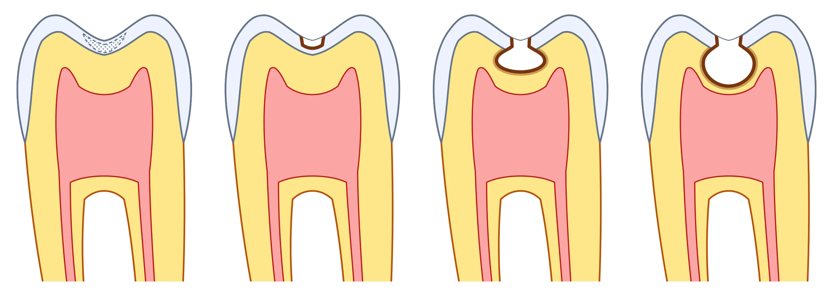
In dentinal caries, the destructive front crosses the enamel-dentin junction (EDJ). Dentin is significantly less mineralized (70% inorganic matter vs. 96% in enamel) and contains dentinal tubules 1–4 µm in diameter, through which acids and bacteria penetrate deeper. In response, odontoblasts form reactive (tertiary) dentin, and hydroxyapatite crystals deposit in the tubules — creating a zone of sclerotic dentin with a protective function.
Histologically, dentinal caries shows: a zone of necrosis (breakdown), a zone of demineralization (soft dentin), a zone of transparent (sclerotic) dentin, and a zone of intact dentin. Pulp vitality is maintained by the intact protective dentin barrier.
Classification
Several caries classifications are used in clinical practice. The most important for understanding dentinal caries are the depth-based classification, the course classification, and Black's topographic classification.
K02.0 Initial caries (white spot lesion) — Lesion limited to the superficial enamel layer. Reversible with timely remineralizing therapy.
K02.0 Enamel caries — Destruction confined to enamel. Carious cavity within the enamel layer, not reaching the EDJ.
K02.1 Dentinal caries — Lesion extends beyond the EDJ into dentin. A layer of intact dentin is preserved between the cavity floor and pulp. Symptoms: brief pain from stimuli.
K02.1 Deep caries — Extensive dentinal involvement with minimal layer over the pulp. High risk of complications. Requires differentiation from pulpitis.
Acute caries — Rapidly progressing form. Dentin is soft, light yellow, easily removed. Often multiple lesions. Typical in young patients with poor oral hygiene.
Chronic caries — Slow progression. Dentin is dense, pigmented (brown or black). More favorable prognosis when treated.
Black's classification (by location) defines the cavity class and accordingly the preparation technique and restorative material selection:
| Class | Location | Affected teeth | Note |
|---|---|---|---|
| I | Pits and fissures of occlusal surfaces | Molars, premolars | Most common location |
| II | Proximal surfaces of posterior teeth | Molars, premolars | Requires matrix when restoring |
| III | Proximal surfaces of anterior teeth (no angle) | Incisors, canines | Esthetically sensitive zone |
| IV | Proximal surfaces with incisal angle | Incisors, canines | Complex angle restoration |
| V | Cervical area (buccal/lingual) | All teeth | High-risk zone (gingival recession) |
| VI | Incisal edge / cusp tip | Incisors, molars | Added later; separate class in some systems |
Clinical Presentation
The clinical presentation of dentinal caries is determined by lesion depth, individual dentin sensitivity, and disease course (acute or chronic).
Brief pain from thermal stimuli — Cold and hot cause pain that resolves immediately after the stimulus is removed. A characteristic symptom.
Pain from chemical stimuli — Sweet, sour, salty foods — also brief pain.
Pain from mechanical stimuli — Food impaction in the cavity, chewing load on the affected tooth.
Aesthetic defect — Visible discoloration or cavity, especially on anterior teeth or fissures.
Food retention in the cavity — Discomfort during chewing, bad breath from decomposing food debris.
Asymptomatic course — In some cases, especially in chronic caries with low dentin sensitivity, complaints may be absent. Diagnosis is made during routine examination.
Objective examination reveals:
| Method | Finding | Note |
|---|---|---|
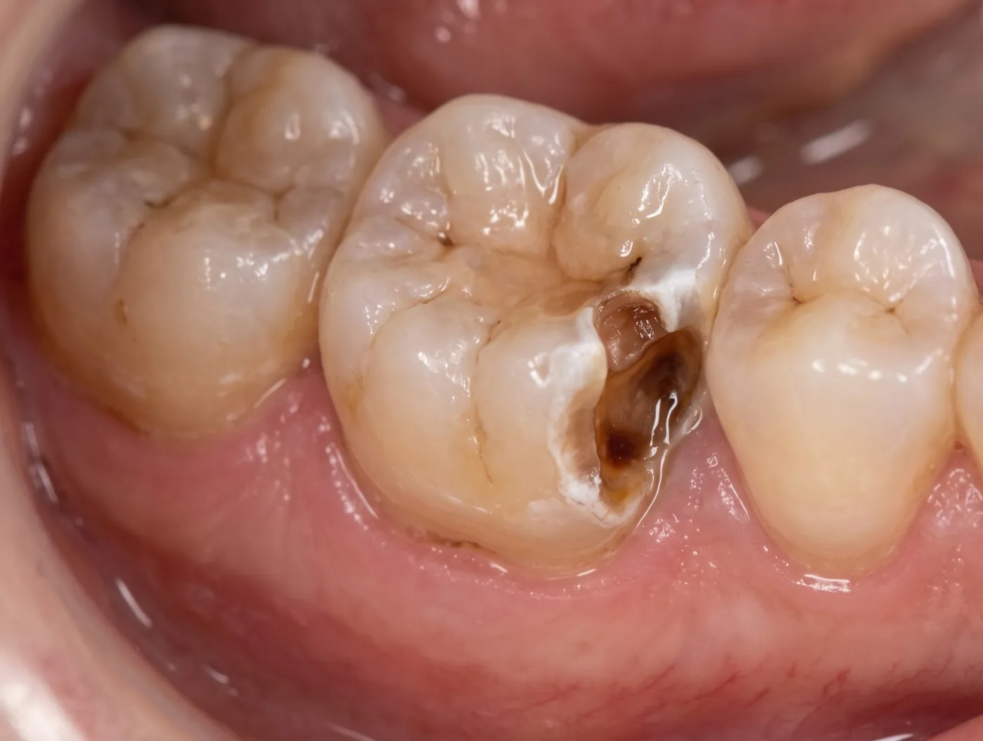
| Inspection | Carious cavity with softened tissues, dark pigmentation | Probe catches without pain or slight pain |
| Probing | Cavity floor painful on probing | Moderate pain within dentin |
| Percussion | Painless | Differentiates from complicated caries |
| Thermal testing | Brief pain from cold | Pain subsides immediately after stimulus removal |
| EPT | 2–12 µA | Pulp is vital (normal 2–6 µA) |
Diagnosis
Diagnosis of dentinal caries is based on a combination of history, objective examination, and additional diagnostic methods. Differential diagnosis is a mandatory step.
| Method | Finding | Note |
|---|---|---|
| Probing | Tenderness along walls and floor within dentin. Dentin may be soft (acute) or dense, pigmented (chronic). | Sharp probe catches in the cavity |
| Vertical percussion | Painless | If painful — rule out periodontitis |
| Horizontal percussion | Painless | |
| Thermal testing (cold) | Brief pain, subsides after stimulus removal | Persistent pain indicates pulpitis |
| Thermal testing (heat) | Mild reaction or normal | Strong heat reaction — sign of pulpitis |
| Electric pulp test (EPT) | 2–12 µA — pulp is vital | Above 100 µA indicates pulp necrosis |
| Radiography | Radiolucent area in dentin not reaching the pulp | Proximal caries — bitewing X-ray mandatory |
| Transillumination | Dark shadow within the crown | Especially useful for anterior teeth |
Treatment
Treatment of dentinal caries includes operative (invasive) and restorative phases. The primary goal is complete removal of necrotic and demineralized tissues, followed by restoration of the anatomical form and function with a restorative material.
Anesthesia — Infiltration or conduction anesthesia. For lower molars — inferior alveolar nerve block. Confirm complete anesthesia after 3–5 minutes. (Articaine 4% + Epinephrine 1:100,000 (Ultracain D-S forte)) (3–5 min)
Field Isolation — Apply rubber dam (OptraDam) to isolate tooth from oral fluids. Mandatory with adhesive systems and composites. Alternative: cotton rolls and saliva ejector. (Rubber dam (OptraDam Plus, Kerr)) (2–3 min)
Cavity Preparation — Remove carious tissues with high-speed handpiece and bur. Sequence: access, extension, necrectomy, shaping. Minimally invasive principle — remove only affected tissues, preserve healthy enamel and dentin. (Burs: round (access), cylindrical, tapered (shaping)) (5–10 min)
Necrectomy Verification — Check removal quality with caries detector dye. Pink-stained areas require additional removal. Floor and walls must be firm and unstained. (Caries Detector (Kuraray) — acid fuchsin-based dye) (1–2 min)
Cavity Disinfection — Rinse with water under pressure and dry. Apply 0.5–2% chlorhexidine gluconate for 30–60 seconds, then dry. Do not use alcohol (denatures dentin proteins). (Chlorhexidine 2% (Vista Dental)) (1 min)
Liner Placement — For deep dentinal lesions — apply calcium-containing liner on cavity floor to protect pulp and stimulate reparative dentin. For dentinal caries, liner may not be required — adhesive only. (Calcium hydroxide (Life, Kerr) or glass ionomer cement (Vitremer, 3M)) (2–3 min)
Etching — Apply 37% phosphoric acid: enamel 15–30 sec, dentin no more than 15 sec (total-etch). After thorough rinsing (10 sec) — gently dry, maintaining dentin moisture ("moist bonding"). (37% Phosphoric acid (Total Etch, Ivoclar / Gel Etchant, Kerr)) (15–30 sec etching + 10 sec rinsing)
Adhesive Application — Apply a one- or two-component adhesive per manufacturer instructions. Actively rub in 15–20 sec, lightly dry, light-cure 20 sec. Forms a hybrid layer with dentin (resin–collagen zone described by Nakabayashi). (Optibond FL (Kerr) or Clearfil SE Bond (Kuraray)) (1–2 min)
Composite Placement and Polymerization — Layer composite ≤2 mm increments, light-cure each layer 20–40 sec. Restore anatomical form considering occlusion. Select shade from Vita scale in natural light. Use stratification for anterior restorations. (Filtek Supreme Ultra (3M) or Charisma Diamond (Kulzer)) (10–15 min)
Finishing and Polishing — Remove excess with finishing burs and discs. Polish with silicone heads and pastes. Check occlusion with articulating paper, adjust contacts. Apply surface sealant (optional). (Sof-Lex discs (3M), Astropol pastes (Ivoclar), BisCover LV (Bisco)) (5–7 min)
Summary
Dentinal caries is one of the most prevalent dental conditions, requiring timely diagnosis and treatment. Understanding pathogenesis mechanisms, clinical presentation, and modern treatment principles enables the clinician to perform high-quality restorations with a long-term prognosis.
The prognosis is favorable when: complete necrectomy is performed, adhesive protocol is correctly followed, polymerization guidelines are observed, and finishing is thorough. Restorations with protocol violations have significantly reduced service life and high risk of secondary caries.
Patients should be informed about prevention: proper brushing technique (Bass or Stillman method), dental floss use, fluoride toothpaste (1450–1500 ppm fluoride), professional hygiene every 6 months.