Inflammation of periapical tissues: forms, diagnosis, radiographic features
ICD-10: K04.4
Definition
Apical (periapical) periodontitis is inflammation of the periodontium in the region of the root apex. It develops as a complication of caries and pulpitis when infection spreads beyond the root canal, or as a consequence of pulp necrosis.
The periodontium is the ligamentous apparatus anchoring the tooth in the alveolus. When inflamed, bone tissue around the root apex is destroyed — visible on radiography as periapical changes.
ICD-10 Code: K04.4–K04.7 — Various forms of periodontitis
EPT: >100 μA — Pulp necrosis — no response
Percussion: Acute: painful — Chronic: often painless
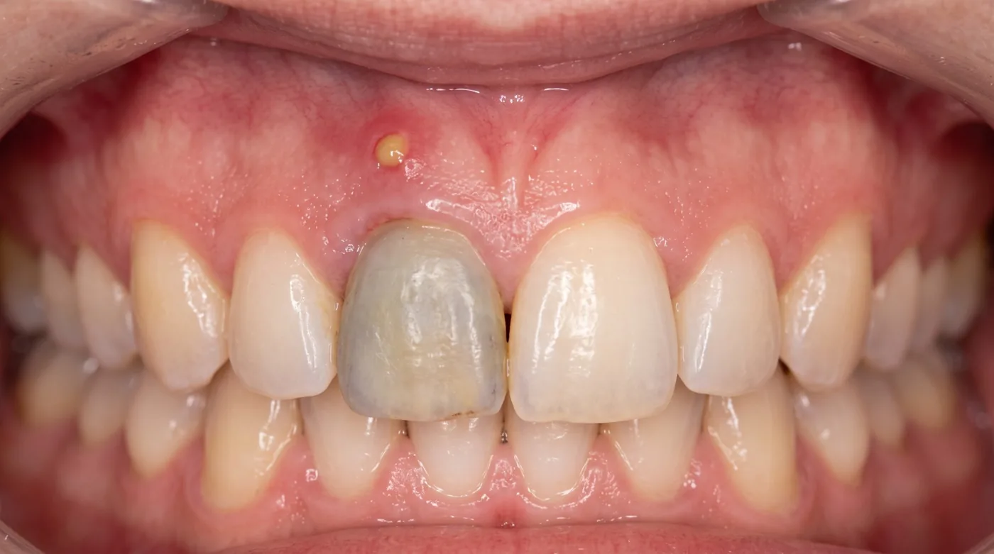
Sinus tract: Granulating — Sinus tract characteristic
X-ray: normal: Sharp line — Uniform periodontal space
X-ray: pathology: Bone lesion — Periapical rarefaction
Classification
K04.4 Acute apical periodontitis (serous) — Initial stage. Progressive pain on biting; tooth feels "elongated." Patient can localize the tooth precisely. Radiograph: widening of the apical periodontal space.
K04.7 Acute apical abscess — Severe throbbing pain, marked soft-tissue swelling. Sharp percussion and palpation pain. Lymphadenopathy, possible fever.
K04.5 Asymptomatic apical periodontitis (legacy chronic fibrous) — Asymptomatic. Found incidentally on X-ray: slight widening of the periodontal space with defined margins at the apex. Outcome of adequately treated tooth or resolved acute episode.
K04.5 Chronic apical periodontitis (legacy granulating) — Actively destructive form. Occasional pain on biting. Characteristic sinus tract on the gingiva with purulent discharge. X-ray: irregular rarefaction with ill-defined "flame-like" margins.
K04.5 Chronic apical periodontitis with granuloma (legacy granulomatous) — Granuloma — rounded bone defect with well-defined margins, surrounded by a fibrous capsule; usually small, most often up to 5–10 mm. Size is only a probabilistic pointer, not a diagnostic criterion: a granuloma cannot be reliably distinguished from a radicular cyst on a radiograph, and the definitive diagnosis is histological. Asymptomatic. Risk of suppuration and cyst formation.
K04.5+ Chronic periodontitis in acute exacerbation — Acute clinical picture superimposed on chronic radiographic changes. Sinus tract may close, increasing pressure and triggering exacerbation.
Clinical Features of Acute Periodontitis
Constant, increasing pain — Unlike pulpitis — pain is continuous, dull-aching, worsening. No pain-free intervals.
"Elongated tooth" sensation — Patient feels the tooth has "grown longer" — due to exudate accumulation in the periodontal space lifting the tooth.
Sharp percussion tenderness — Light tapping causes acute pain — "positive percussion." This is the cardinal clinical sign of acute periodontitis.
Pain on biting and contact — Patient avoids occluding. Any contact with the opposing tooth causes sharp pain.
Soft-tissue swelling (in abscess) — Inflammatory infiltrate and collateral oedema of the cheek or lip, lymphadenitis. Possible fever.
Radiographic Features
| Form | Radiographic appearance | Margin character |
|---|---|---|
| Acute serous | Widening of the periodontal space at the apex | Well-defined |
| Chronic fibrous | Slight uniform widening of the space | Well-defined, smooth |
| Chronic granulating | Irregular bone rarefaction ("flame" pattern) | Ill-defined, diffuse |
| Chronic granulomatous | Rounded rarefaction at the apex, most often up to 5–10 mm | Well-defined, rounded (capsule) |
| Cystgranuloma / root cyst | Rounded defect, most often >10 mm, with corticated border | Very sharp, "punched-out" |
Differential Diagnosis
| Feature | Acute periodontitis | Symptomatic irreversible pulpitis (diffuse; legacy acute diffuse) | Periostitis |
|---|---|---|---|
| Pain character | Constant, worsening | Paroxysmal | Constant, diffuse |
| Percussion | Sharply painful | Mildly + or painless | Painful |
| EPT | >100 μA (necrosis) | 15–35 μA | >100 μA |
| Swelling | In abscess | None | Marked, in vestibular fold |
| Fluctuation | None | None | Present in mature abscess |
| X-ray | Widened space / periapical lesion | Normal or slight widening | Cortical plate intact |