Fluorosis, hypoplasia, wedge-shaped defect, erosion — classification and differential diagnosis
ICD-10: K00.3
Definition and Classification
Non-carious lesions of hard dental tissues are a group of pathological conditions in which enamel and dentin are destroyed without microbial involvement — fundamentally distinguishing them from caries.
In Kazakhstan and Russia, the Patrikeev classification (1968) is used, dividing lesions into those arising before tooth eruption (systemic) and after eruption (local).
Classification by time of onset
Before eruption (systemic enamel formation disorders): Enamel hypoplasia (K00.40), Dental fluorosis (K00.30–K00.39), Tetracycline staining, Hereditary disorders: amelogenesis imperfecta, dentinogenesis imperfecta
After eruption (action on the erupted tooth): Wedge-shaped defect (K03.1), Tooth erosion (K03.2), Excessive attrition / pathological tooth wear (K03.0), Dentinal hypersensitivity (K03.8), Abfraction, Acid necrosis (K03.2)
Dental Fluorosis
Fluorosis is a systemic enamel condition caused by excessive fluoride intake during tooth formation (before age 6–8). Fluoride disrupts ameloblast function, producing abnormal enamel mineralization.
Endemic fluorosis occurs when drinking water contains more than 1.5 mg/L of fluoride (optimal range 0.8–1.2 mg/L). Kazakhstan has endemic fluorosis regions.
Questionable / Very mild (streaky) — Barely visible chalky striations on the labial surface of incisors. Very mild fluorosis.
Mild (macular) — Milky-white opaque spots without defined margins. Surface smooth. Spots may coalesce giving a whitish appearance.
Moderate (chalky-pitted) — Yellow-brown staining and pitting of enamel. Moderate fluorosis.
Severe (erosive) — Large brown stains with surface erosion; enamel tissue lost. Severe fluorosis.
Very severe (destructive) — Crown destruction. Seen in highly endemic areas with fluoride >6 mg/L.
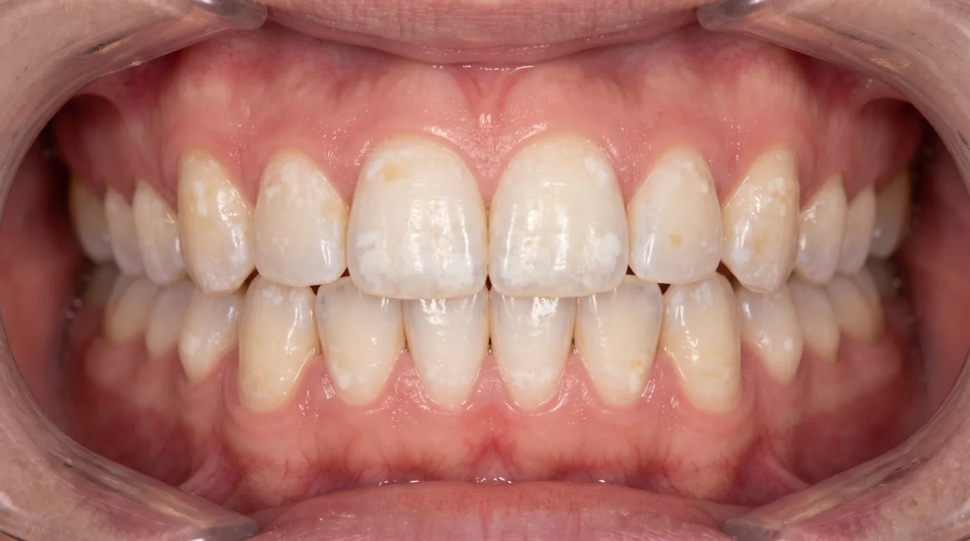
Enamel Hypoplasia
Hypoplasia is underdevelopment of enamel due to metabolic disturbance during its formation. Caused by systemic (systemic hypoplasia) or local (local hypoplasia) factors acting on ameloblasts.
| Method | Finding | Note |
|---|---|---|
| Systemic hypoplasia | Symmetrical defects on homologous teeth. Grooves, pits, or spots at the same crown level (chronological marking) | Causes: childhood illness in first year, rickets, maternal toxemia |
| Local hypoplasia (Turner tooth) | Affects one or a few permanent teeth — usually premolars. White spots, grooves, or cup-shaped pits | Causes: periapical abscess of primary predecessor, trauma |
| Hutchinson incisors | Notched cutting edge on upper central incisors, barrel-shaped crown | Sign of congenital syphilis |
| Pflüger / Moon molars | Convergent crown form on first molars ("bud" or "mulberry" molar) | Sign of congenital syphilis |
Wedge-Shaped Defect and Erosion
Wedge-shaped defect (K03.1) — Hard-tissue loss in the cervical region with two walls converging at 40–45°. Surface smooth, hard, glossy. Location: canines and premolars. Dentinal hypersensitivity. Cause: horizontal brushing technique, abrasive toothpaste, parafunctions.
Tooth erosion (K03.2) — Saucer-shaped hard-tissue loss on the labial surface. Surface smooth, glazed. Bilateral symmetrical. Cause: acids (GERD, citrus, fizzy drinks, bulimia).
Excessive attrition / pathological tooth wear (K03.0) — Hard-tissue loss of occlusal and incisal surfaces. Grades: I (enamel), II (dentin to 2/3 crown height), III (>2/3), IV (to cervix). Cause: bruxism, abrasive diet, hard toothbrushes.
Abfraction — Cervical wedge-shaped defect caused by occlusal stress. Distinguished from classic wedge defect by triangular form, acute apical angle, often subgingival location.
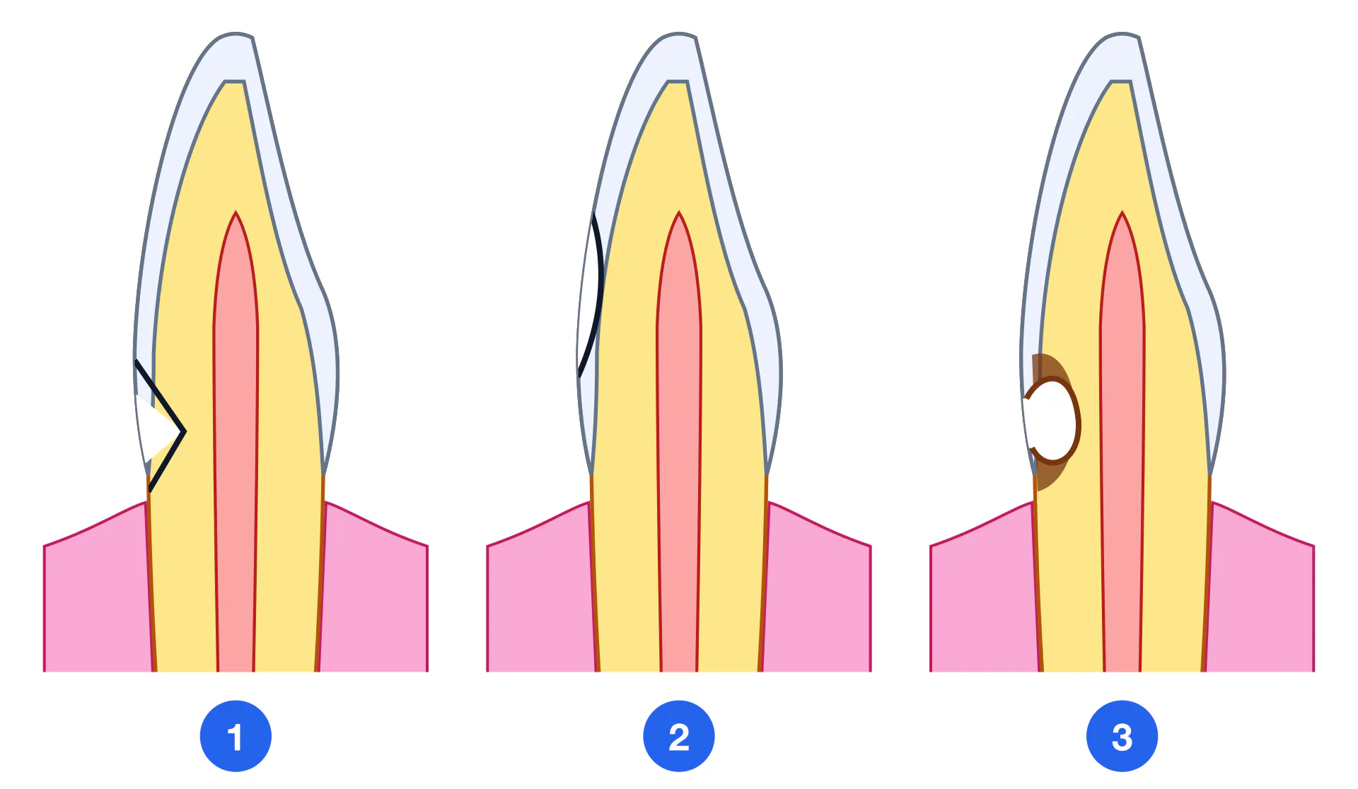
| Feature | Wedge-shaped defect | Erosion | Cervical caries |
|---|---|---|---|
| Shape | Wedge, 40–45° | Saucer / cup | Cavity with undermined margins |
| Surface | Smooth, glossy | Smooth, mirror-like | Rough, soft |
| Location | Cervical, labial/buccal | Labial, coronal third | Often proximal or occlusal |
| Bilateral | Often bilateral | Yes, bilateral | No |
| Progression | Slow | Moderate | Active |
| Probing | Painless, hard | Painless, hard | Painful, soft dentin |
Differential Diagnosis
| Feature | Fluorosis (macular) | Initial caries (spot) | Hypoplasia |
|---|---|---|---|
| Time of onset | Before eruption | After eruption | Before eruption |
| Symmetry | Symmetrical | Asymmetrical | Symmetrical (systemic) |
| Spot color | Chalky or yellow-brown | Chalky/brown | White or yellow |
| Enamel lustre | Dull / chalky | Loss of lustre | Preserved or dull |
| Methylene blue stain | Negative | Positive | Negative |
| Surface texture | Smooth | Rough, chalky | Smooth or pitted |